PAPER NOTES

Paper notes

Read the evidence. Bring it back to the bench.

Read cell death and inflammation research through experimental questions, original paper figures and related antibody testing notes.

Preparing search…

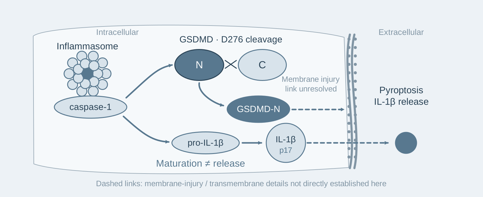
Inflammasome caspase-1 drives IL-1β maturation and GSDMD cleavage at D276 separately. The N-terminal fragment mediates pyroptosis and release, with membrane-injury details shown as unresolved.
PAPER NOTE · 01

After GSDMD knockout, caspase-1 is still activated and IL-1β still matures, yet the cells no longer undergo pyroptosis

Introduction: Why do cells swell and burst after an inflammasome activates caspase-1? Before 2015, this question had no answer: none of the known caspase-1 substrates at the time could explain pyroptosis. The authors knocked out the 9 proteins identified by their screen one by one. Only 1 knockout prevented cell death and IL-1β release: GSDMD.

Cell Research · 2015 / 8 min read

Read the paper note
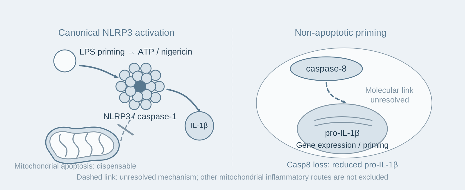
At left, canonical NLRP3 activation by ATP/nigericin after LPS priming does not require mitochondrial apoptosis. At right, caspase-8 contributes to pro-IL-1β priming without apoptosis; the intervening molecular link is unresolved.
PAPER NOTE · 02

After testing 14 gene-knockout mouse strains one by one, none supported the popular model of "mitochondrial apoptosis activating NLRP3"

Introduction: Around 2012, the most popular explanation in the field was that diverse NLRP3 stimuli first induce mitochondrial apoptosis, and the released mitochondrial components then activate NLRP3. However, this model relied mainly on inhibitor experiments and observations of correlation, without gene-knockout tests. This study tested 14 gene-knockout mouse strains one by one, and none supported the model; the real new finding concerned another molecule—caspase-8 has a function that does not promote cell death.

EMBO Reports · 2014 / 9 min read

Read the paper note
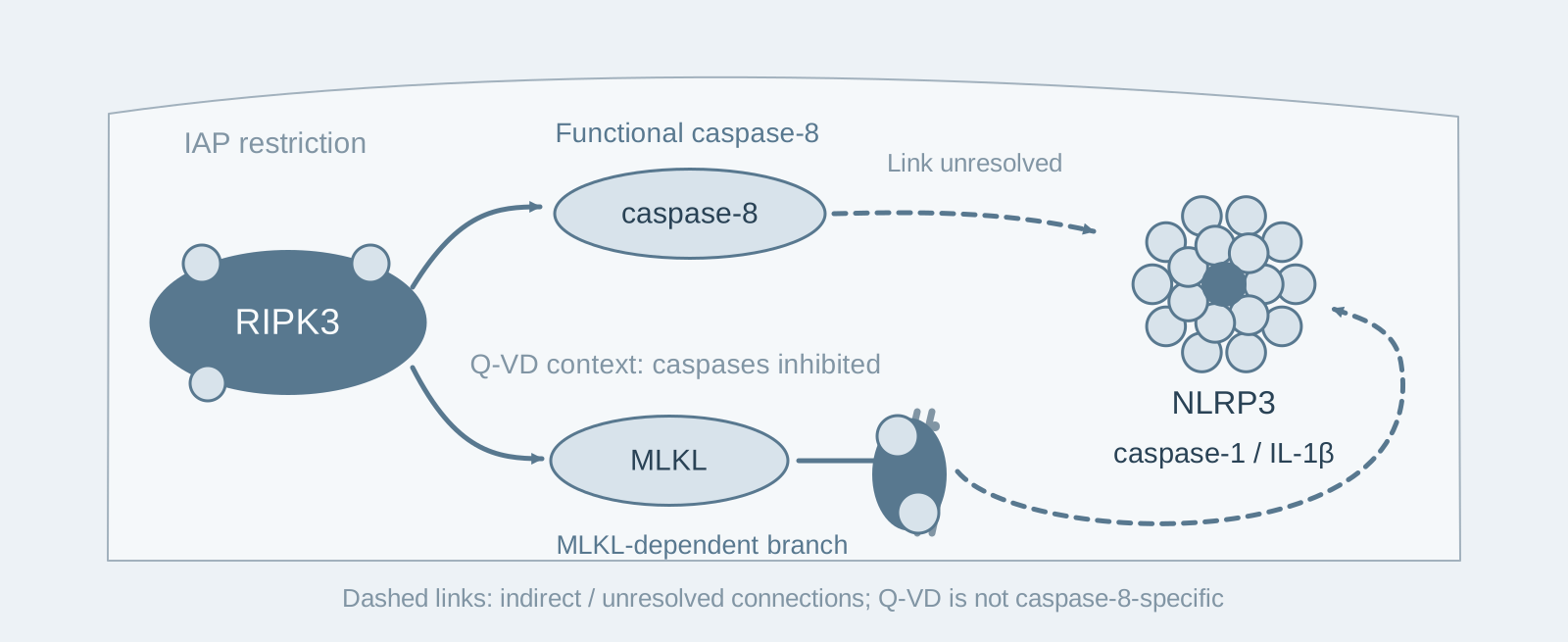
When caspase-8 is functional, RIPK3 engages NLRP3 through a caspase-8-associated, MLKL-independent branch. Under Q-VD conditions, an MLKL-dependent branch predominates; unresolved downstream links are dashed.
PAPER NOTE · 03

Deleting MLKL does not block RIPK3's pro-inflammatory activity: RIPK3 activates NLRP3 through caspase-8

Introduction: RIPK3's pro-inflammatory activity is generally thought to require its substrate MLKL, indirectly activating the inflammasome through necroptosis. In macrophages lacking MLKL, this study found that caspase-1 cleavage and IL-1β secretion induced by LPS plus an IAP inhibitor remained completely normal, whereas deleting RIPK3 caused a marked defect—RIPK3 can activate NLRP3 without MLKL.

Nature Communications · 2015 / 9 min read

Read the paper note
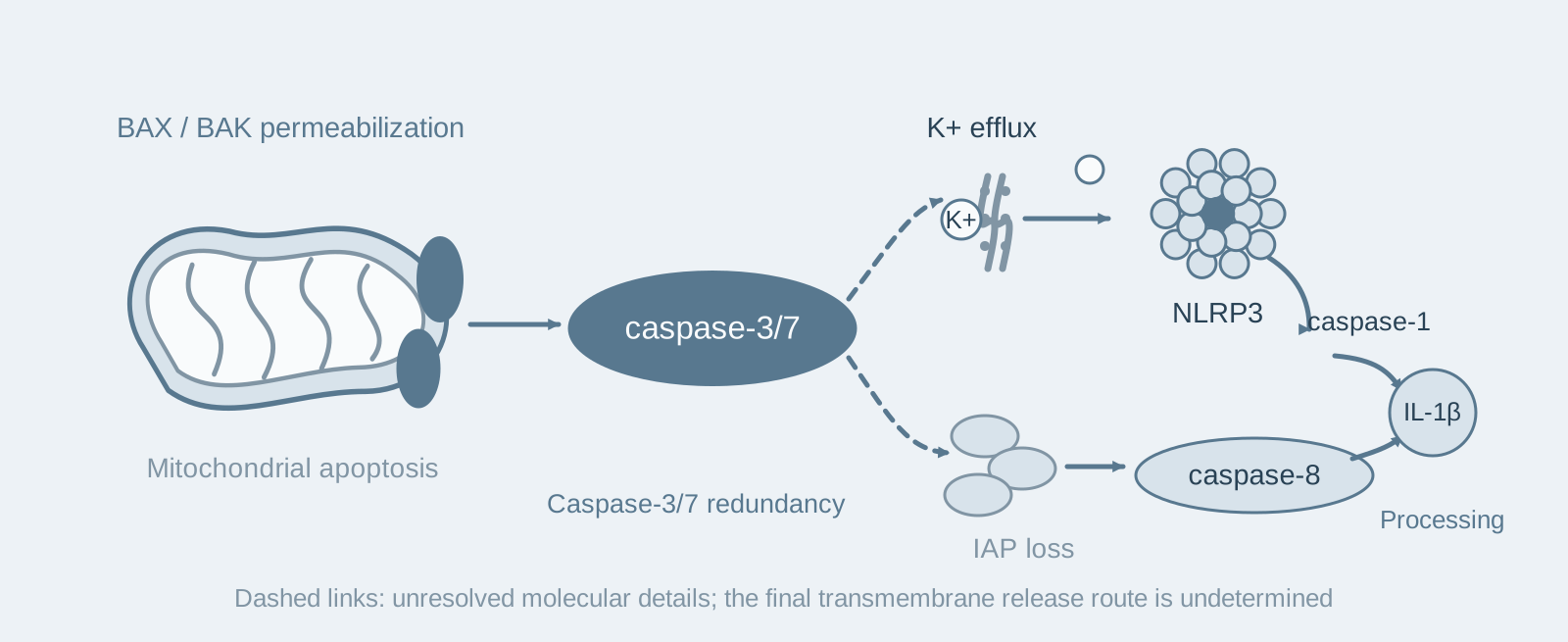
After BAX/BAK-mediated mitochondrial permeabilization, caspase-3/7 engage a potassium-efflux/NLRP3/caspase-1 branch and a parallel IAP-loss/caspase-8 branch, both processing IL-1β.
PAPER NOTE · 04

Mitochondrial apoptosis, considered "immunologically silent," can also activate IL-1β: BAX/BAK engages two pro-inflammatory pathways simultaneously through caspase-3/7

Introduction: Mitochondrial apoptosis is usually considered an "immunologically silent" mode of cell death: the cells do not rupture their membranes and are promptly cleared by phagocytes. This paper finds that once BAX/BAK permeabilizes the mitochondrial outer membrane, apoptotic macrophages release IL-1β, while TNF secretion remains completely unaffected at the same time. What is silent is the morphology of apoptosis, rather than its inflammatory consequences.

Cell Reports · 2018 / 8 min read

Read the paper note
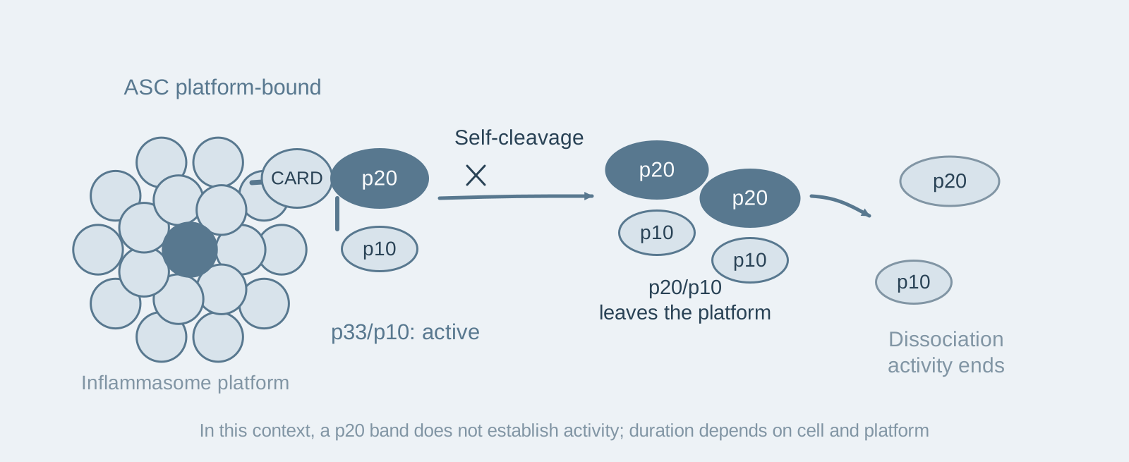
Active ASC-platform-bound p33/p10 retains its CARD-containing linkage. After self-cleavage, p20/p10 leaves the platform and dissociates, terminating activity in the stated cellular context.
PAPER NOTE · 05

The caspase-1 p20 band on a Western blot is actually an inactive product: how self-cleavage terminates inflammasome activity

Introduction: Textbooks describe the p20/p10 tetramer produced by caspase-1 self-cleavage after inflammasome activation as the active form. The authors used an activity probe to directly capture active caspase-1 in cells and found that the main species captured was p33/p10, with almost no p20—p20/p10 is not the active product, but a remnant left after caspase-1 inactivation.

Journal of Experimental Medicine · 2018 / 8 min read

Read the paper note
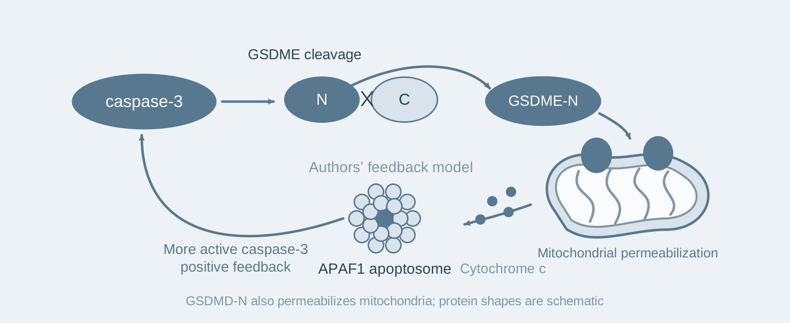
Caspase-3 cleavage generates GSDME-N, which permeabilizes mitochondria and releases cytochrome c. The APAF1 apoptosome amplifies caspase-3 and yields more GSDME-N.
PAPER NOTE · 06

Gasdermin pores are not limited to the plasma membrane: the N-terminal fragments of GSDME and GSDMD can also permeabilize mitochondria

Introduction: The authors deleted GSDME in human leukemia cells, initially expecting only a reduction in pyroptosis. Instead, caspase-3 activation itself became slower and weaker. A recognized downstream substrate of caspase-3 was influencing caspase-3 activation in return—this paper shows why: besides the plasma membrane, GSDME-N targets a second membrane compartment, the mitochondria.

Nature Communications · 2019 / 8 min read

Read the paper note
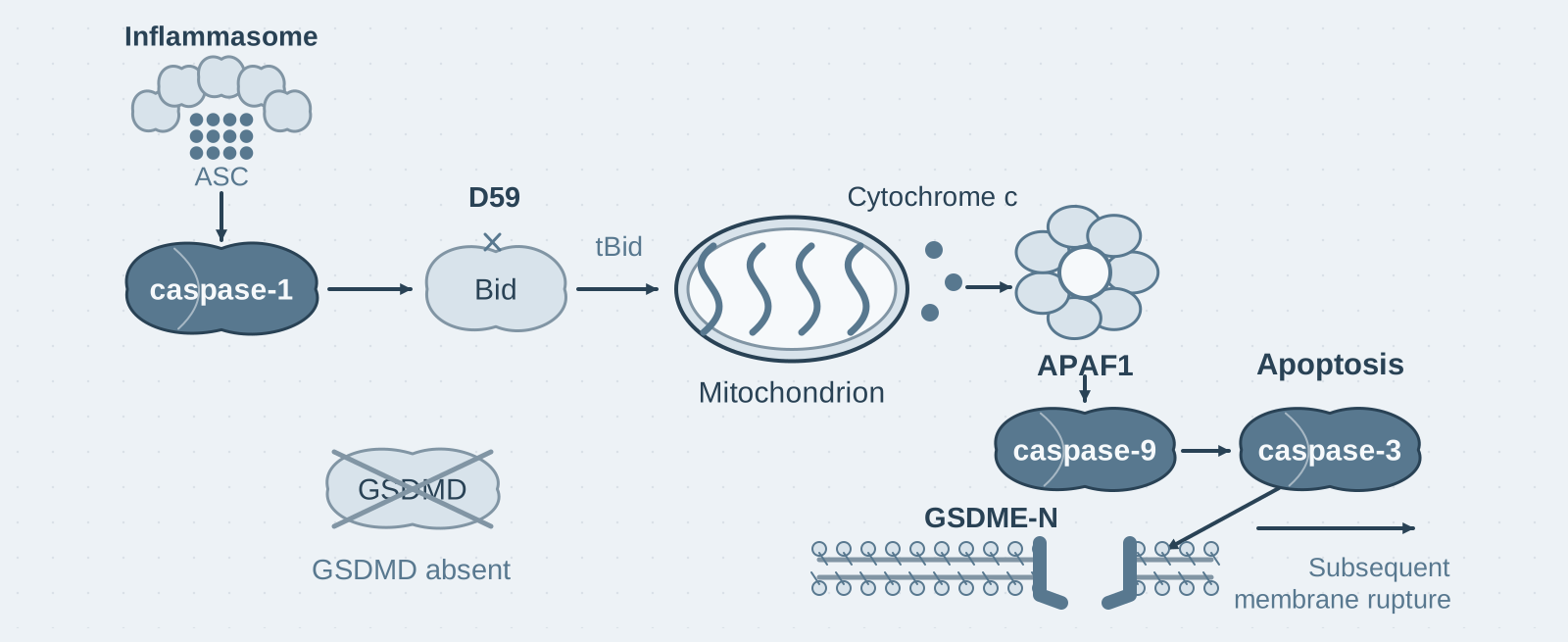
In GSDMD-deficient cells, caspase-1 cleaves Bid at D59, engaging cytochrome c, APAF1 and caspase-9 to activate caspase-3; GSDME can contribute to subsequent membrane rupture. Only the principal Bid-dependent route is depicted.
PAPER NOTE · 07

Inflammasomes remain functional in the absence of GSDMD: caspase-1 cleaves Bid to initiate apoptosis

Introduction: After Salmonella infection, Gsdmd-knockout macrophages do not undergo the typical rapid pyroptosis, but they still die. Death is slower, the morphology shifts to an apoptosis-like pattern with membrane blebbing, and caspase-3, -8, and -9 are all activated. What caspase-1 does when it loses GSDMD as a substrate is the question this paper addresses.

Nature Communications · 2019 / 8 min read

Read the paper note
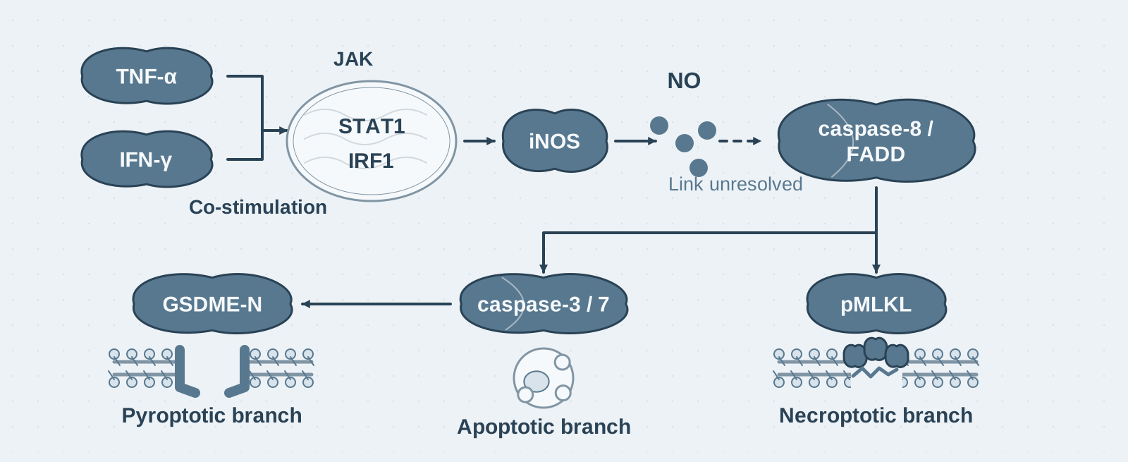
TNF-α and IFN-γ engage JAK/STAT1/IRF1–iNOS/NO signalling and caspase-8/FADD-dependent PANoptosis. GSDME, caspase-3/7 and pMLKL branches are shown in parallel; the molecular link between NO and caspase-8 remains unresolved and is dashed.
PAPER NOTE · 08

The only pair among 28 cytokine combinations that kills macrophages: how TNF-α + IFN-γ triggers PANoptosis

Introduction: More than a dozen inflammatory factors are elevated simultaneously in the serum of patients with severe COVID-19, but which ones cause disease has remained unresolved. The authors treated macrophages with all pairwise combinations of 8 candidate factors. Of the 28 combinations, only TNF-α and IFN-γ killed the cells; neutralizing these two factors in mice significantly reduced mortality in models of cytokine shock, sepsis, HLH, and SARS-CoV-2 infection.

Cell · 2021 / 10 min read

Read the paper note
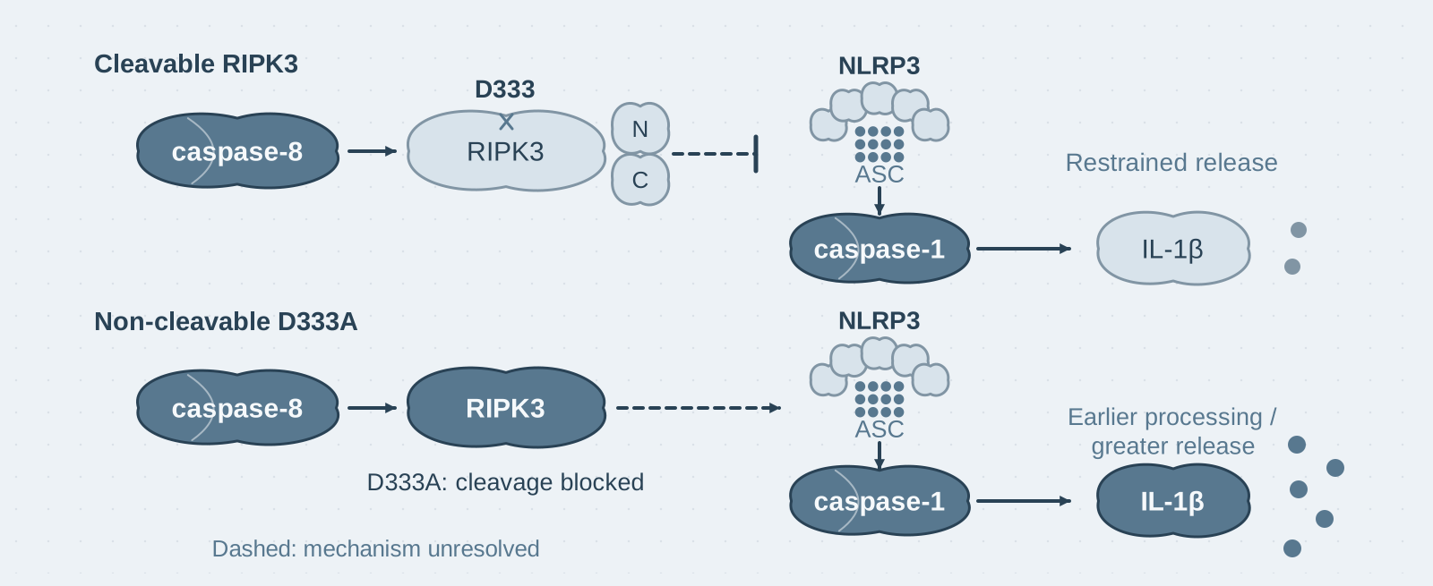
Under TLR priming and IAP inhibition, caspase-8 cleavage of RIPK3 at D333 restrains NLRP3 activation, caspase-1 processing and IL-1β release. The non-cleavable D333A mutant enhances these inflammatory readouts. The dashed inhibitory link summarises observations rather than a resolved molecular mechanism.
PAPER NOTE · 09

A cleavage site strictly conserved through evolution, yet mice remain healthy after it is mutated: RIPK3 cleavage does not govern necroptosis

Introduction: The textbook model holds that caspase-8 prevents necroptosis by cleaving RIPK3. The authors of this study mutated this cleavage site, yet the mice remained healthy for more than 400 days. The site's actual role lies in another pathway: when IAPs are inhibited, it provides negative feedback that limits the intensity of NLRP3 inflammasome activation.

Cell Death & Differentiation · 2024 / 8 min read

Read the paper note
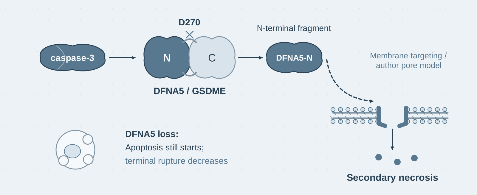
Caspase-3 cleaves DFNA5 (GSDME) at D270. Its N-terminal fragment targets the plasma membrane and enables secondary necrosis following apoptosis. The pore is a schematic author model rather than a structure resolved in this paper; DFNA5 loss does not prevent apoptosis initiation.
PAPER NOTE · 10

Membrane rupture at the end of apoptosis is also programmed: caspase-3 cleaves DFNA5 to execute secondary necrosis

Introduction: If apoptotic cells are not cleared promptly, they eventually swell, lose membrane integrity, and release their contents. This secondary necrosis was long regarded as passive osmotic disintegration. The authors began with an observation: macrophages lacking inflammatory caspases through a double knockout still swelled and ruptured after viral infection. Testing human caspases one by one, they found that only caspase-3 could cleave DFNA5, and that the resulting N-terminal fragment was responsible for forming pores in the cell membrane.

Nature Communications · 2017 / 9 min read

Read the paper note
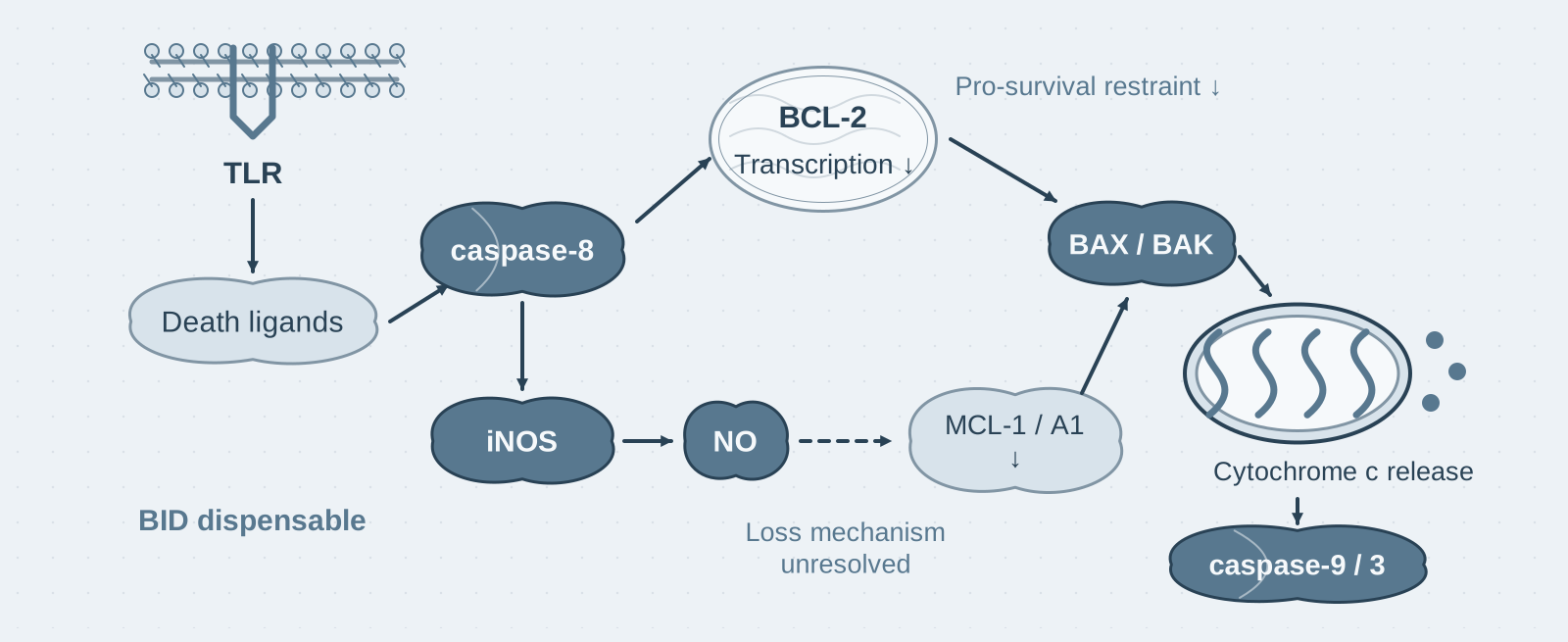
After IFN-γ priming and subsequent TLR stimulation, death ligands and caspase-8 engage BAX/BAK-mediated mitochondrial death. Reduced BCL-2 transcription and iNOS/NO-dependent loss of MCL-1 and A1 relieve pro-survival restraints. The molecular mechanism of NO-dependent protein loss is unresolved and dashed; BID is dispensable.
PAPER NOTE · 11

IFN-γ alone does not kill macrophages, but TLR ligands given after priming do: how caspase-8 and mitochondrial apoptosis connect infection to cell death

Introduction: IFN-γ is a classic macrophage-activating factor and does not kill cells when given alone. This paper found that macrophages primed with IFN-γ overnight and then exposed to TLR ligands such as LPS died within 24 hours; replacing IFN-γ with IFN-β for priming caused no death at all. Death was executed by caspase-8 and the BAX/BAK mitochondrial apoptosis pathway, while nitric oxide produced by iNOS was a prerequisite for activation of this pathway.

Immunity · 2022 / 10 min read

Read the paper note
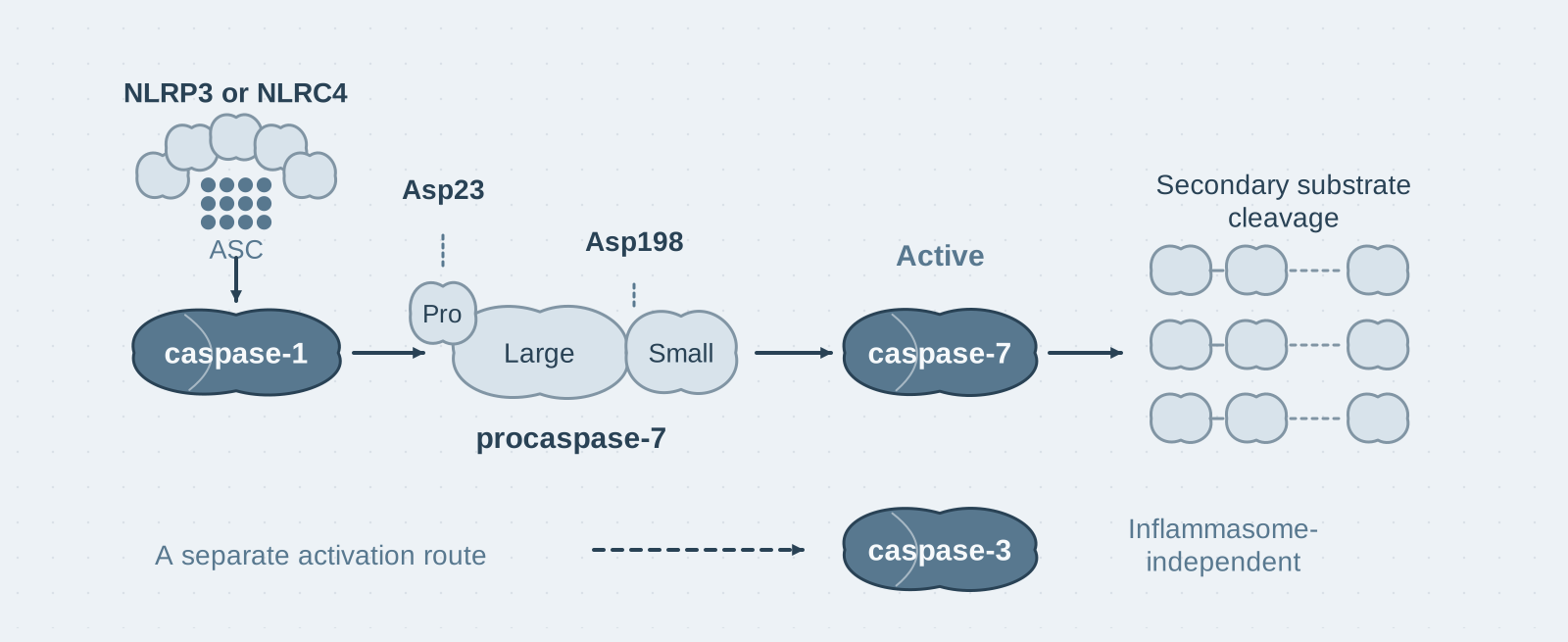
The NLR–caspase-1 inflammasome cascade cleaves procaspase-7 at Asp23 and Asp198, generating active caspase-7 and secondary substrate cleavage. Caspase-3 activation can proceed independently of the inflammasome. This processing cascade does not establish caspase-7 as necessary for infection-induced death.
PAPER NOTE · 12

A proteomics screen reveals that the caspase-1 inflammasome directly cleaves and activates caspase-7

Introduction: The authors used proteomics to screen macrophage lysates for caspase-1 substrates. Of 1022 proteins, only 20 were cleaved after Asp—one of them was caspase-7, which "executes apoptosis", and its cleavage site was precisely its canonical activation site. More unexpectedly, half of the cleavage events identified looked less like the work of caspase-1 and more like that of caspase-7.

Molecular & Cellular Proteomics · 2008 / 9 min read

Read the paper note
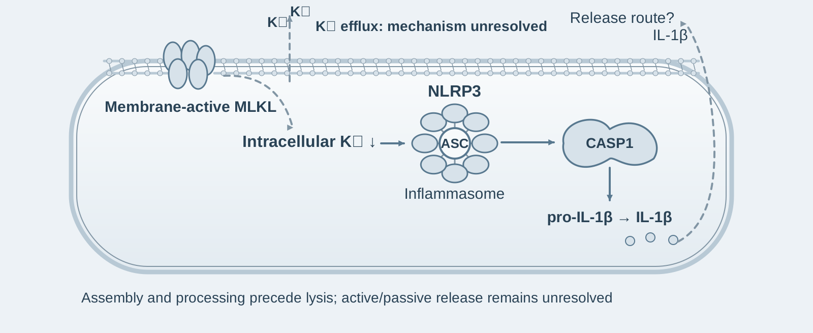
Independent blue-gray mechanism schematic: membrane-active MLKL induces potassium efflux, linking reduced intracellular potassium to NLRP3–ASC assembly and caspase-1-dependent IL-1β processing. The efflux arrow is separated from MLKL subunits and its specific mechanism is marked unresolved. A dashed arrow and question mark mark the unresolved IL-1β release mechanism.
PAPER NOTE · 13

The inflammation of necroptosis is not "leaked out": active MLKL activates NLRP3 through potassium efflux before cell rupture

Introduction: The textbook explanation for inflammation caused by necroptosis is that cellular contents (DAMPs) leaking out after cell rupture stimulate surrounding cells. In this study, the authors suppressed cell lysis with the osmoprotectant glycine and found that IL-1β was still secreted—inflammation was actively generated by MLKL activating NLRP3 before cell rupture, rather than passively leaking out afterward.

Proceedings of the National Academy of Sciences of the United States of America · 2017 / 8 min read

Read the paper note
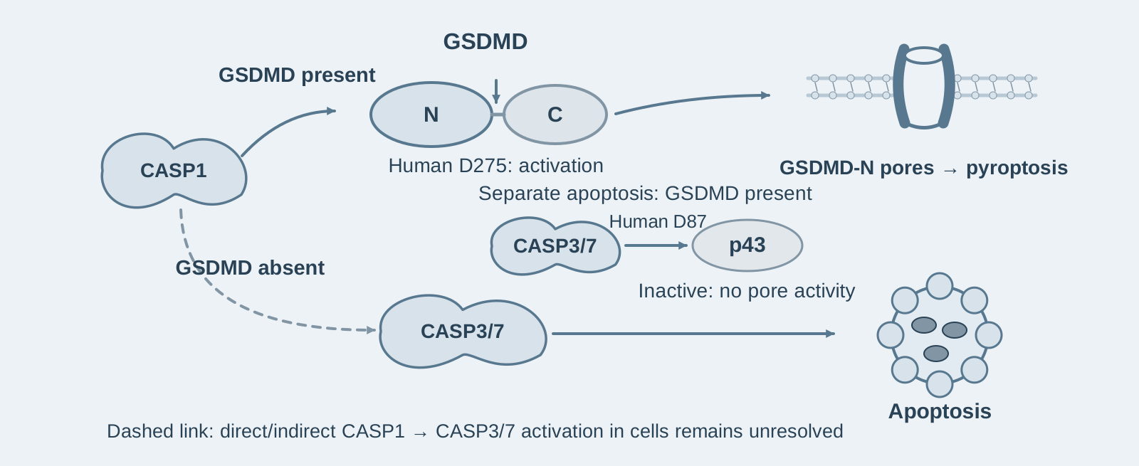
Independent blue-gray schematic: caspase-1 cleavage of human GSDMD at D275 produces a pore-forming N terminus. Without GSDMD, signaling activates caspase-3/7 and apoptosis, with its unresolved direct or indirect cellular link shown dashed. A separate branch explicitly represents apoptosis with GSDMD present, where caspase-3/7 cleavage at human D87 inactivates GSDMD and yields p43.
PAPER NOTE · 14

Bidirectional crosstalk between pyroptosis and apoptosis: without GSDMD, caspase-1 triggers apoptosis, while apoptotic caspases inactivate GSDMD in turn

Introduction: The same caspase-1 activation leads to pyroptosis when GSDMD is present; after GSDMD knockout, cells instead undergo apoptosis. In turn, once apoptosis begins, caspase-3/7 cleave GSDMD at another site (D87) to produce an inactive p43 fragment, actively preventing pyroptosis. Crosstalk between the two death pathways is bidirectional, with opposite directions.

Cell Chemical Biology · 2017 / 8 min read

Read the paper note
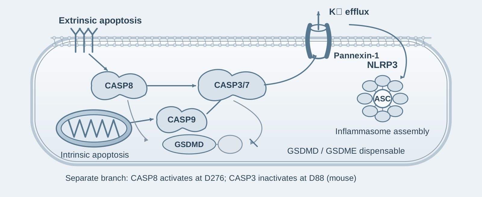
Independent blue-gray schematic: extrinsic caspase-8 and mitochondrial intrinsic caspase-9 pathways converge on caspase-3/7, connecting membrane pannexin-1 activity and potassium efflux to NLRP3 assembly. A separate branch shows mouse GSDMD activation at D276 by caspase-8 and inactivation at D88 by caspase-3; gasdermin pores are not drawn as the upstream NLRP3 channel.
PAPER NOTE · 15

Apoptosis is not immunologically silent: caspase-8 directly cleaves GSDMD to cause cell lysis, while pannexin-1 drives NLRP3 activation

Introduction: Apoptosis has long been regarded as an "immunologically silent" form of cell death. This study, however, finds that extrinsic apoptosis initiated by caspase-8 activation cleaves GSDMD into pore-forming p30 and causes cell lysis. When the caspase-3 inactivation site D88 in GSDMD is mutated, the mice are born healthy, but their macrophages show a 1.5-fold increase in cell death during apoptosis. Apoptosis normally avoids becoming pyroptosis because caspase-3 inactivates GSDMD by cleaving it.

The EMBO Journal · 2019 / 9 min read

Read the paper note
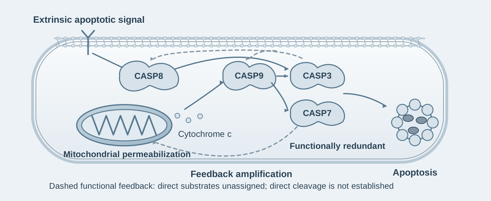
Independent blue-gray schematic: extrinsic caspase-8 and mitochondrial/caspase-9 signals activate redundant caspase-3 or caspase-7 and apoptosis. Curved dashed arrows return to initiator caspases and mitochondria, representing experimentally supported feedback amplification without claiming identified direct cleavage substrates.
PAPER NOTE · 16

Effector caspases are more than executors: caspase-3/-7 amplify upstream apoptotic signals through feedback

Introduction: Simultaneously knocking out effector caspase-3 and -7 in leukemia cells reduces their sensitivity to apoptosis-inducing agents by more than four orders of magnitude. The reason is not simply that the "executioners" are gone: activation of upstream initiator caspase-8 and -9, and mitochondrial permeabilization itself, require feedback amplification by downstream effector caspases.

Science Advances · 2019 / 9 min read

Read the paper note
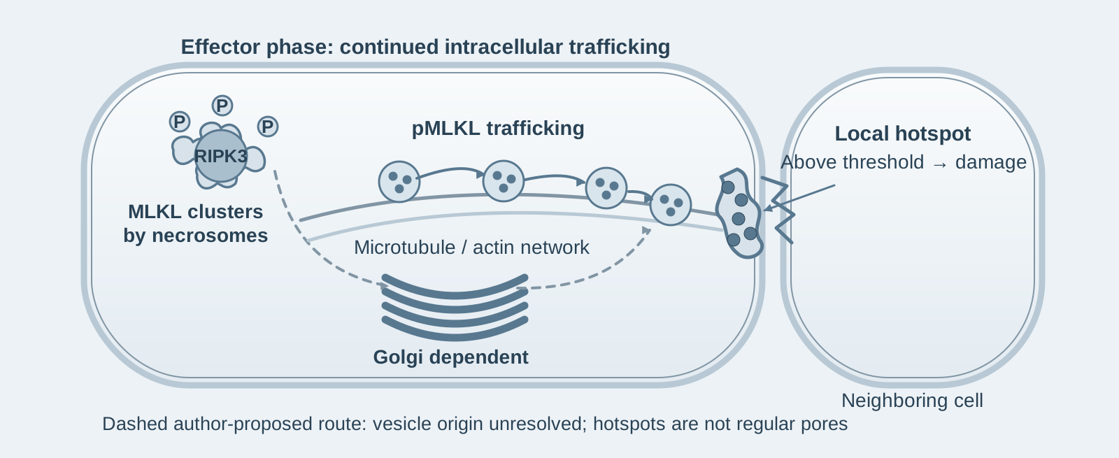
Independent blue-gray mechanism schematic: phosphorylated MLKL clusters near cytoplasmic necrosomes, then accumulates at plasma-membrane hotspots near cell contacts through Golgi-, microtubule- and actin-dependent trafficking, preceding local damage. The proposed Golgi-derived vesicle route is dashed; irregular hotspots are not depicted as regular protein pores.
PAPER NOTE · 17

MLKL phosphorylation does not immediately rupture the membrane: intracellular trafficking determines the speed and threshold of necroptosis

Introduction: Cells do not die immediately after MLKL is phosphorylated—in HT29 cells, this delay exceeds 3 hours. This study finds that the intervening period is used to transport phosphorylated MLKL from the necrosome, through Golgi, microtubule and actin networks, to 1–2 hotspots on the plasma membrane. Membrane rupture occurs only when MLKL at these hotspots exceeds a threshold; membrane damage is also passed to adjacent cells, accelerating necroptosis in their neighbors by approximately 25%.

Nature Communications · 2020 / 8 min read

Read the paper note
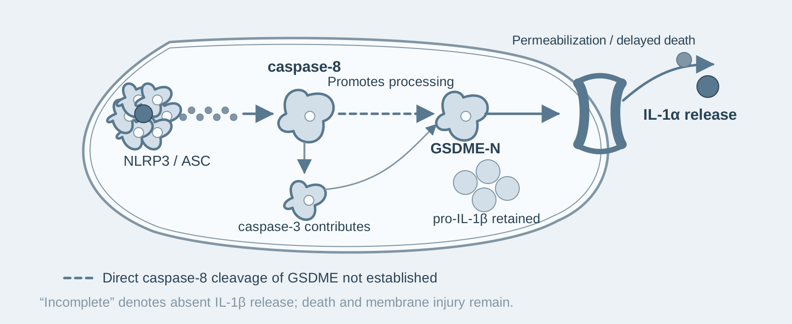
Blue-gray schematic of the tested NLRP3 activation model under caspase-1 loss or inhibition: ASC and caspase-8 promote GSDME processing, delayed death and IL-1α release, while pro-IL-1β is retained. A dashed connection explicitly marks unproven direct caspase-8 cleavage of GSDME.
PAPER NOTE · 18

An alternative death program after caspase-1 inhibition: caspase-8 switches to cleaving GSDME, permitting IL-1α release through "incomplete pyroptosis"

Introduction: According to the textbook account, knocking out caspase-1/11 should block inflammasome-mediated pyroptosis—but this study found that, after nigericin activates NLRP3, wild-type macrophages begin to die at 40 minutes, whereas caspase-1/11 double-knockout cells still die after 180 minutes, simply using a different set of execution machinery. More unusually, this form of death fails to release IL-1β but still releases IL-1α.

iScience · 2020 / 10 min read

Read the paper note
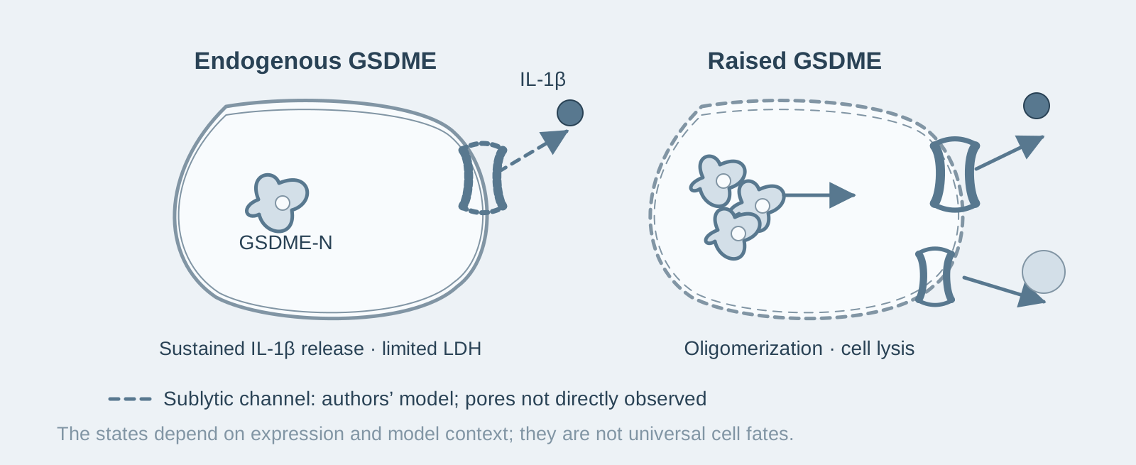
Blue-gray comparison in the GSDMD-deficient models: endogenous GSDME supports IL-1β release with limited LDH, consistent with the authors’ inferred sublytic channel model; raised GSDME promotes oligomerization and lysis. A dashed channel denotes pores not directly observed in the sublytic state.
PAPER NOTE · 19

GSDME expression determines the outcome: endogenous levels sustain IL-1β release without lysing cells, whereas only high expression leads to pyroptosis

Introduction: After inflammasome activation in GSDMD-knockout macrophages, IL-1β continues to be released for at least 5 hours, while the cell membrane remains largely unruptured—the executor is GSDME at endogenous levels. Yet the same GSDME causes complete pyroptosis once it is overexpressed. Its expression level determines which path it takes.

Cell Reports · 2021 / 9 min read

Read the paper note
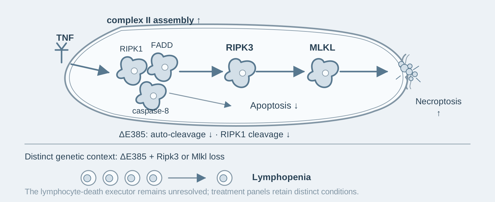
Blue-gray schematic under TNF-related stimuli: caspase-8 ΔE385 reduces auto-cleavage and RIPK1 cleavage, enhances complex II-related signaling, reduces apoptosis and favors RIPK3–MLKL necroptosis. A separate lower branch shows lymphopenia with combined Ripk3 or Mlkl loss in mice; its executor remains unresolved.
PAPER NOTE · 20

Loss of caspase-8 auto-cleavage: weaker apoptosis, enhanced necroptosis, and lymphopenia

Introduction: Mice with a complete knockout of caspase-8 die during embryonic development; in this study, deleting just one amino acid (E385) in its auto-cleavage site allowed mice to survive to adulthood. Yet the loss of this single amino acid was sufficient to switch TNF-induced apoptosis to necroptosis and cause severe lymphopenia on a Ripk3- or Mlkl-knockout background.

Cell Death & Differentiation · 2022 / 10 min read

Read the paper note
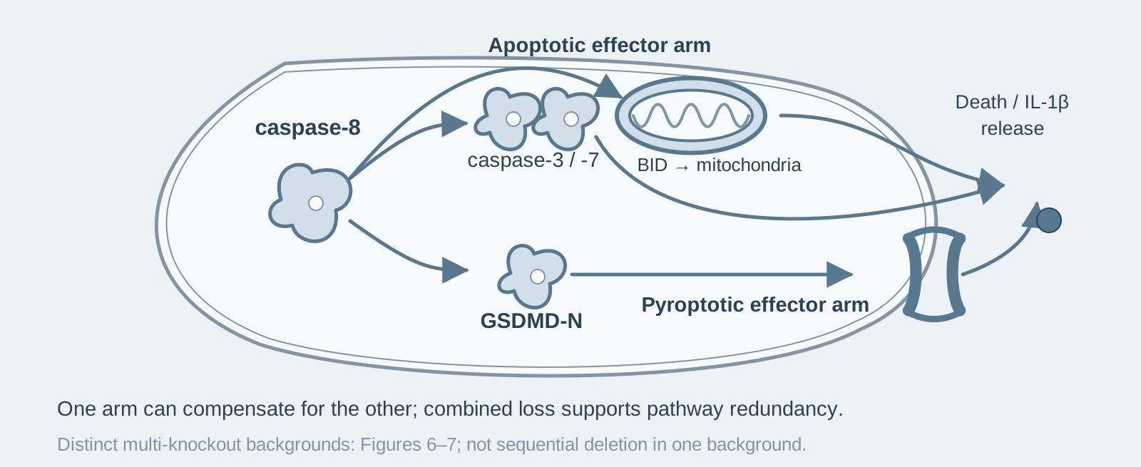
Blue-gray schematic of the authors’ redundancy model in IAP-targeted macrophages: apoptotic effectors and the GSDMD pyroptotic arm downstream of caspase-8 can compensate for each other to support death and IL-1β release. Combined-loss genotypes support the model; distinct multi-knockout backgrounds are not depicted as sequential isogenic deletion.
PAPER NOTE · 21

Apoptotic and pyroptotic machinery are redundant in XIAP deficiency: only combined deletion blocks cell death and IL-1β release

Introduction: Macrophages lacking 7 cell-death execution molecules (caspase-1, -3, -7, -11 and -12, BID and MLKL) still die in response to XIAP inhibitors, as efficiently as wild-type cells. Complete protection is achieved only when GSDMD is additionally deleted. This study thereby demonstrates that the apoptotic and pyroptotic machinery downstream of caspase-8 are redundant.

The EMBO Journal · 2023 / 10 min read

Read the paper note
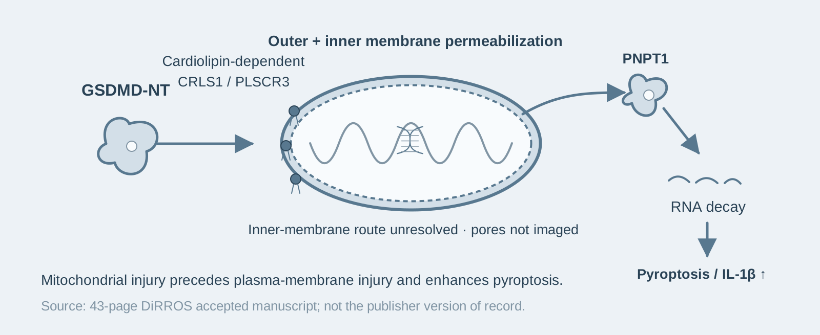
Blue-gray schematic: GSDMD-NT binding to cardiolipin supports permeabilization of mitochondrial outer and inner membranes, releasing PNPT1 and other contents and promoting RNA degradation and enhanced pyroptosis. Mitochondrial injury precedes plasma-membrane injury; pores were not directly imaged and the inner-membrane mechanism remains unresolved. Based on the 43-page accepted manuscript.
PAPER NOTE · 22

GSDMD breaches both mitochondrial membranes before the plasma membrane: mitochondrial damage accelerates and amplifies pyroptosis

Introduction: During pyroptosis, GSDMD has already breached both the outer and inner mitochondrial membranes before it forms pores in the plasma membrane. This study finds that such mitochondrial damage is not a by-product of death: knocking out the cardiolipin synthase CRLS1 or PLSCR3, which transports cardiolipin to the outer mitochondrial membrane, markedly inhibits both pyroptosis and IL-1β release.

Immunity · 2023 / 8 min read

Read the paper note