- 01
Main finding: The proteomics screen identified 20 substrates cleaved downstream of caspase-1, including caspase-7;
- 02
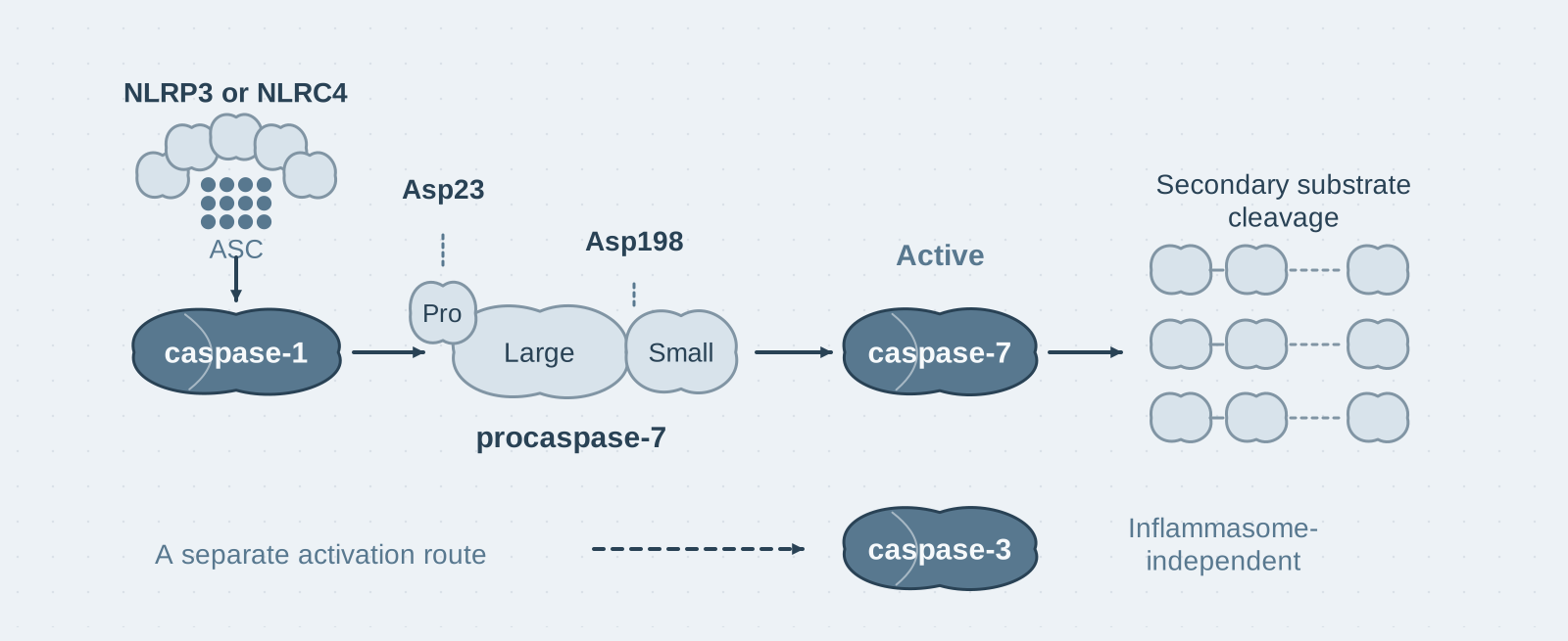
Key mechanism: Caspase-1 cleaves procaspase-7 at Asp23 and Asp198 to activate it; under inflammatory stimulation, caspase-7 activation depends on the caspase-1 inflammasome, whereas caspase-3 activation does not;
- 03
Significance: Provides the first demonstration of an NLR–caspase-1–caspase-7 cascade and reveals a divergence between the mechanisms that activate caspase-3 and caspase-7 under inflammatory stimulation.
On this page
I. What this paper answers
What else does caspase-1 cleave besides the precursors of IL-1β and IL-18? This paper answers through a differential proteomics screen: the 20 candidate substrates include the effector caspase-7, which caspase-1 directly cleaves at its canonical activation sites and activates. Under inflammatory conditions in vivo, caspase-7 activation depends entirely on the caspase-1 inflammasome, whereas caspase-3, another effector caspase, takes a separate route that does not depend on the inflammasome.
II. The logic of the evidence
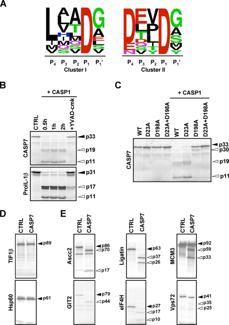
Step 1: A COFRADIC screen identifies caspase-7 and exposes "cascade contamination" (Figure 1A, Tables I–II). The authors combined SILAC labelling with COFRADIC (a proteomics method that enriches protein N-terminal peptides and can precisely locate cleavage sites) to compare macrophage lysates treated with recombinant caspase-1 against untreated lysates: 2663 peptides corresponded to 1022 proteins, of which 20 were specifically cleaved after Asp (23 sites in total). Analysis divided the cleavage sites into two groups: 11 (48%) resembled the caspase-1 cleavage site in pro-IL-1β (a hydrophobic aliphatic residue at P4); caspase-7 was in this group, with the site IQAD198 between its large and small subunits. The remaining 12 (52%) instead resembled the DEVD sequence preferred by caspase-7. These results indicate that the added caspase-1 activated endogenous caspase-7 in the lysate, and half of the cleavage events identified were produced by a secondary caspase.
Step 2: In vitro validation that caspase-1 cleaves caspase-7 at its canonical activation sites (Figure 1B–E). The authors incubated 35S-labelled procaspase-7 with 30 nM recombinant caspase-1 and found that the 33 kDa precursor was completely processed into the active p19/p11 subunits within 30 minutes; the caspase-1 inhibitor YVAD-cmk blocked this entirely. Point mutations mapped cleavage to Asp23 (removal of the prodomain) and Asp198 (between the large and small subunits); mutating both sites prevented processing. Recombinant caspase-7 could cleave the second group of substrates (Assc2, GIT2, Ligatin, eIF4H, MCM3 and Vps72), but could not cleave the first group even at a 10-fold concentration. These results show that caspase-1 directly cleaves and activates caspase-7, and the second group of substrates is indeed produced indirectly by caspase-7.
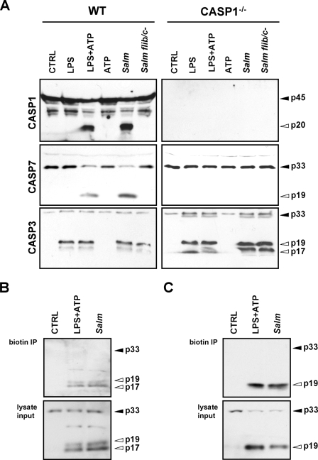
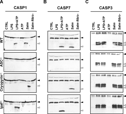
Step 3: In vivo genetic evidence—caspase-7 activation depends on the caspase-1 inflammasome, whereas caspase-3 activation does not (Figures 2–5). The authors tested bone marrow-derived macrophages from a series of knockout mice: LPS + ATP or Salmonella infection could activate caspase-7, but this activation disappeared completely in caspase-1- or ASC-deficient cells. Salmonella-induced caspase-7 processing required flagellin and Ipaf (now called NLRC4), whereas LPS + ATP required Cryopyrin (now called NLRP3). Caspase-3, however, was activated as usual in all of the knockout cells (LPS alone was sufficient). Pull-down experiments with a biotin-labelled DEVD-fmk probe confirmed the presence of activated caspase-3 (p17/p19) and caspase-7 (p19). These results indicate that under inflammatory stimulation, caspase-7 activation is driven by the NLR–caspase-1 inflammasome, while caspase-3 follows an independent route.
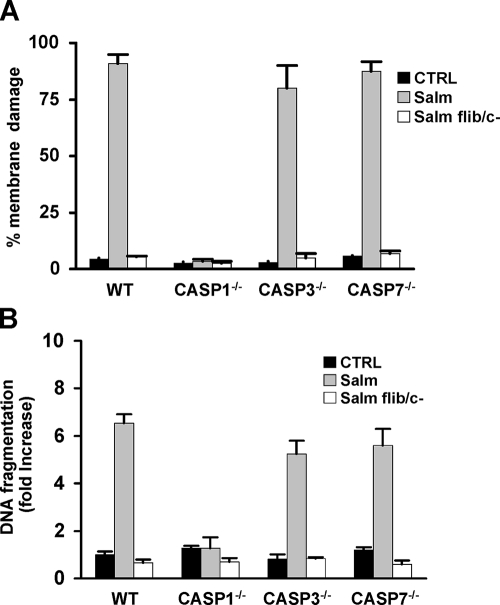
Step 4: The two routes are nonredundant, and neither is the executor of infection-induced death (Figures 3 and 6). Knocking out caspase-3 did not affect activation of caspase-1 or caspase-7, and knocking out caspase-7 did not affect caspase-1 or caspase-3. Macrophages lacking either caspase-3 or caspase-7 were not protected from Salmonella-induced death. These results indicate that the mechanisms activating caspase-3 and caspase-7 are nonredundant in this model, and neither is individually a necessary executor of infection-induced death.
III. Implications for experiments
- Guard against "cascade contamination" in degradomics screens: Direct substrates and substrates of secondary caspases become mixed together, so cleavage-site motif analysis must first separate them into groups, followed by individual validation—in this paper, 52% of the events were actually produced by caspase-7.
- Caspase-7 activation can serve as a downstream readout of caspase-1 inflammasome activity: Under inflammatory stimulation, caspase-7 processing depends on caspase-1, while caspase-3 processing does not—the two bands convey different information and cannot be used interchangeably.
- DEVD probes and inhibitors do not distinguish the two: Biotin-DEVD-fmk labels both caspase-3 and caspase-7; "DEVDase activity" or a phenotype caused by DEVD inhibition therefore cannot be attributed to a single caspase.
IV. Links to other papers on this site
- Extension | Caspase-1 self-cleavage terminates inflammasome activity (Boucher 2018, J Exp Med): Caspase-1 cleavage events have two different functions—this paper shows that caspase-1 cleaves caspase-7 at Asp23/Asp198 and activates it (signal transmission), while Boucher shows that caspase-1 self-cleavage produces p20, causing it to dissociate from the inflammasome and become inactive (signal termination).
- Extension | Bidirectional crosstalk between pyroptosis and apoptosis (Taabazuing 2017, Cell Chem Biol): This paper establishes a cascade in which caspase-1 directly cleaves and activates caspase-7; Taabazuing demonstrates that, when GSDMD is absent, caspase-1 activation of caspase-3/7 redirects cells from pyroptosis to apoptosis—the cascade described here provides its biochemical basis.
- Comparison | Effector caspases amplify apoptotic signalling through feedback (McComb 2019, Sci Adv): This paper finds that downstream of the inflammasome, caspase-7 activation depends on caspase-1 whereas caspase-3 activation does not (divergent mechanisms); McComb demonstrates that caspase-3/-7 functions are fully redundant in apoptosis—the relationship between the same pair of effector caspases differs across pathways.
(The comparisons across papers above are integrative interpretations, rather than direct conclusions of any one original paper.)
What remains unresolved
Limitations: ① The screen was performed in lysates, and the added 400 nM caspase-1 exceeds physiological concentrations, so the substrate repertoire may include sites specific to in vitro conditions; ② this paper does not establish the physiological function of caspase-7 after inflammasome activation (it neither protects against nor worsens infection-induced death), and the significance of its downstream substrates remains for later investigation.
Targeted Peptidecentric Proteomics Reveals Caspase-7 as a Substrate of the Caspase-1 Inflammasomes
Mohamed Lamkanfi; Thirumala-Devi Kanneganti; Petra Van Damme; Tom Vanden Berghe; Isabel Vanoverberghe; Joël Vandekerckhove; Peter Vandenabeele; Kris Gevaert; Gabriel Núñez Molecular & Cellular Proteomics. 2008;7:2350-2363.
The original PDF was supplied by the user, verified against its title, authors, DOI and complete figures, and stored without modification. The verified original figures retain their labels, axes and controls. The blue-gray cover is an independently drawn mechanism schematic with separate source records.
References
- Lamkanfi M, Kanneganti TD, Van Damme P, et al. Targeted peptidecentric proteomics reveals caspase-7 as a substrate of the caspase-1 inflammasomes. Mol Cell Proteomics. 2008;7(12):2350-2363. doi:10.1074/mcp.M800132-MCP200
- Mariathasan S, Newton K, Monack DM, et al. Differential activation of the inflammasome by caspase-1 adaptors ASC and Ipaf. Nature. 2004;430(6996):213-218. doi:10.1038/nature02664
Paper discussion
No sign-up. Nickname optional.