- 01
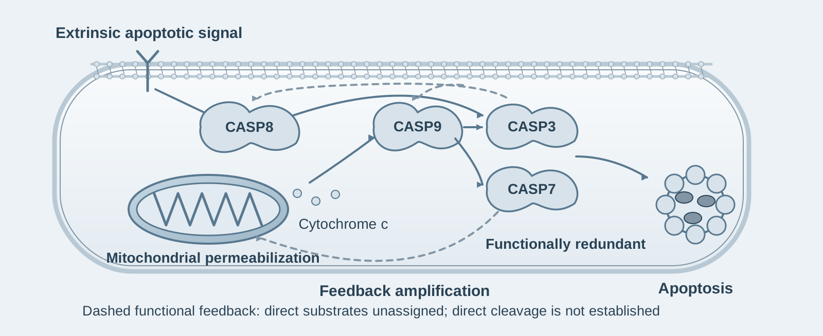
Main finding: Caspase-3 and -7 are functionally redundant. Their combined knockout makes leukemia cells highly resistant to extrinsic and intrinsic apoptosis-inducing agents, whereas caspase-6 has no evident role;
- 02
Key mechanism: Without effector caspases, upstream caspase-8/-9 activation is delayed and weakened, mitochondrial membrane potential is maintained, and cytochrome c release is delayed—effector caspases amplify upstream signals through feedback;
- 03
Significance: The apoptotic cascade is not unidirectional. "Downstream" effector caspases determine whether "upstream" events can be completed fully, explaining developmental lethality in double-knockout mice.
On this page
1. What question does this paper answer?
The classical model of the apoptotic cascade is unidirectional: initiator caspases (-8 and -9) activate effector caspases (-3, -7, and -6), which execute cell death. This paper asks: How much does each of the three effector caspases contribute in human leukemia cells? Can upstream events still occur normally when they are absent? Its conclusion is that caspase-3 and -7 are completely redundant: either one alone can execute apoptosis, whereas loss of both produces high resistance. Caspase-6 makes no measurable contribution. More significantly, activation of upstream caspase-8/-9 and mitochondrial permeabilization are both markedly impaired in cells lacking caspase-3/-7. Efficient completion of the apoptotic cascade therefore requires feedback amplification by effector caspases.
2. Following the chain of evidence
Step 1: Both extrinsic and intrinsic apoptotic stimuli activate caspase-8 and -9 together (Figs. 1–2). In NALM6 leukemia cells, the authors induced extrinsic apoptosis with Birinapant, a Smac mimetic, plus TNF, and intrinsic apoptosis with ABT263, a BCL-2 family inhibitor. Flow-cytometric FLICA activity measurements showed that both stimuli activated caspase-8 and -9 together. Gene knockout confirmed their classical division of roles: caspase-8 knockout protected cells against Bir/TNF but not ABT263, whereas caspase-9 knockout partially protected against ABT263 but not Bir/TNF. Changes in mitochondrial membrane potential, measured by TMRE, corresponded to these results. Together, these findings indicate that the two pathways initiate separately but both lead to activation of both initiator caspases.
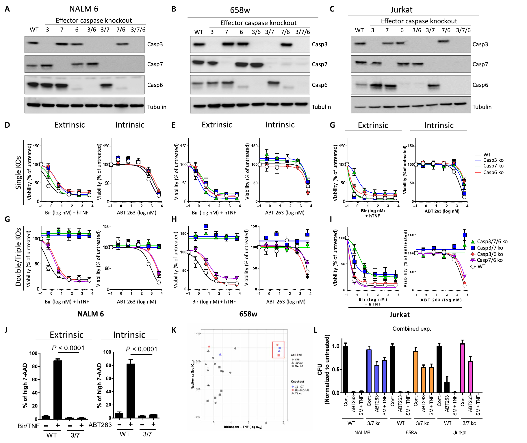
Step 2: Caspase-3/-7 execute apoptosis redundantly, whereas caspase-6 has no role (Fig. 3). The authors used CRISPR to knock out caspase-3, -7, and -6 individually or in combination in three leukemia cell lines: NALM6, patient-derived 658w, and Jurkat. Individual knockouts had little effect on drug sensitivity. Combined caspase-3/7 knockout increased the IC50 for Bir/TNF by more than four orders of magnitude in NALM6 and 658w cells, and colony-formation assays confirmed long-term survival. Jurkat was the exception: double-knockout cells resisted ABT263 but remained sensitive to Bir/TNF because Jurkat cells switch to necroptosis when apoptosis is blocked. Additional caspase-6 knockout provided no further protection. Together, these results indicate that caspase-3 and -7 redundantly execute apoptosis, whereas caspase-6 is not an essential executor.
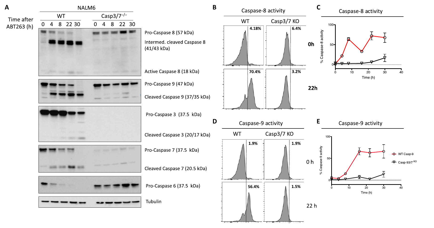
Step 3: Loss of effector caspases weakens activation of upstream initiator caspases (Figs. 4–5). Repeating both apoptosis inductions in caspase-3/7 double-knockout cells, the authors found that cleavage and activity of caspase-8 and -9 were markedly delayed and reduced. Caspase-9 activation was almost abolished. Together, these results indicate that full activation of upstream initiator caspases depends on feedback amplification by downstream effector caspases: the cascade is not unidirectional.
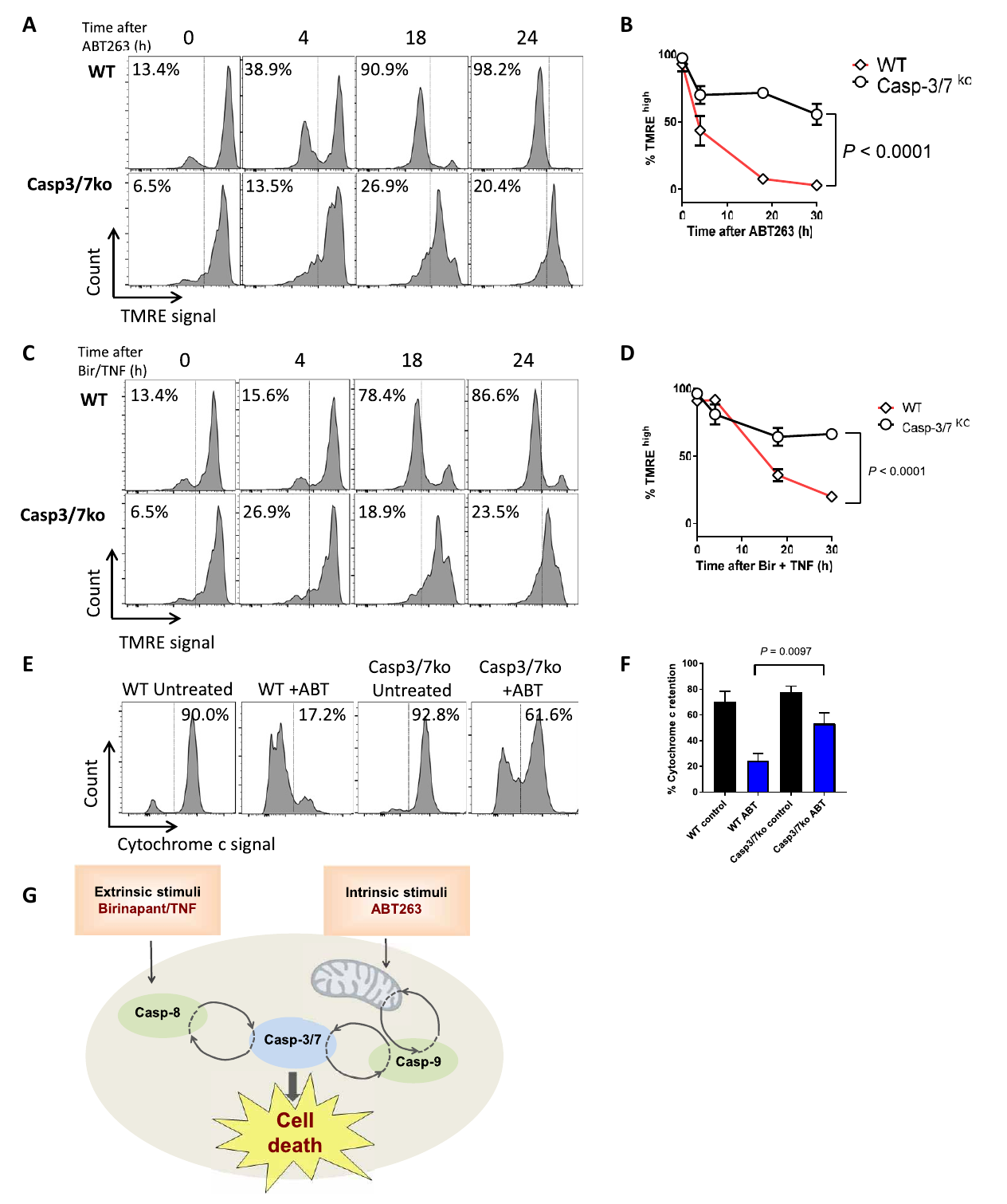
Step 4: Effector caspase activity is required for mitochondrial depolarization and cytochrome c release (Fig. 6). The authors measured mitochondrial membrane potential and cytochrome c release. Wild-type cells completely depolarized within 24 hours after apoptosis induction, whereas double-knockout cells largely maintained their membrane potential and showed delayed cytochrome c release. Together, these results indicate that effector caspase feedback extends upstream to mitochondria, contributing to completion of mitochondrial permeabilization.
3. Implications for experiments
- Low initiator caspase activity does not mean the pathway has not initiated: In the absence of effector caspases, FLICA readouts and cleavage blots for caspase-8/-9 drop markedly. Feedback amplification must be considered as a confounding factor when assessing the extent of upstream activation.
- Both caspase-3 and -7 must be knocked out to reveal the phenotype: They are completely redundant, so a negative result from a single knockout does not rule out an effector caspase role. Caspase-6 knockout can serve as a control to test for the absence of an additional effect.
- Blocking apoptosis in hematological tumor cells may redirect death toward necroptosis: This explains why Jurkat double-knockout cells remain sensitive to Bir/TNF. The mode of death should be assessed alongside drug resistance.
4. Connections to other papers on this site
- Extension | Gasdermins permeabilize mitochondria to amplify caspase-3 (Rogers 2019, Nat Commun): Positive feedback in apoptotic signaling operates at two levels. This paper shows that effector caspases amplify upstream caspase-8/-9 and mitochondrial permeabilization through protein cleavage and feedback. Rogers 2019 shows that GSDME-N permeabilizes mitochondria to amplify caspase-3 activation itself.
- Extension | Redundant apoptotic and pyroptotic machinery in XIAP deficiency (Hughes 2023, EMBO J): This paper shows that caspase-3/-7 are completely redundant in executing apoptosis. Hughes et al. show that redundancy extends beyond the effector caspases themselves: in XIAP deficiency, the apoptotic and pyroptotic machineries serve as backups for one another.
- Comparison | Caspase-1 inflammasomes activate a caspase-7 cascade (Lamkanfi 2008, Mol Cell Proteomics): This paper shows complete functional redundancy of caspase-3/-7 in apoptosis. Lamkanfi et al. find that caspase-7 activation downstream of inflammasomes depends on caspase-1, whereas caspase-3 activation does not. The same pair of effector caspases therefore has different relationships in different pathways.
- Comparison | Caspase-8 self-cleavage regulates the apoptosis/necroptosis switch (Li 2022, Cell Death Differ): Both papers describe reduced apoptotic efficiency, but the mechanisms act at different positions. Here, loss of effector caspase-3/-7 attenuates upstream signals. In Li et al., loss of initiator caspase-8 self-cleavage weakens apoptosis.
(The comparisons above are integrated interpretations across papers, rather than direct conclusions of any single original paper.)
What remains unresolved
Limitations: ① The direct molecular substrates of feedback amplification were not identified. The authors suggest that it may involve effector caspases cleaving XIAP to relieve inhibition; ② All experiments were conducted in leukemia cell lines, with no validation in primary cells or in vivo.
Efficient apoptosis requires feedback amplification of upstream apoptotic signals by effector caspase-3 or -7
Scott McComb; Pik Ki Chan; Anna Guinot; Holmfridur Hartmannsdottir; Silvia Jenni; Maria Pamela Dobay; Jean-Pierre Bourquin; Beat C. Bornhauser Science Advances. 2019;5:.
Figures are from the verified original paper stored in the library. Original labels, axes and controls are retained; figure numbers and sources are registered in the library.
References
- McComb S, Chan PK, Guinot A, et al. Efficient apoptosis requires feedback amplification of upstream apoptotic signals by effector caspase-3 or -7. Sci Adv. 2019;5(7):eaau9433. doi:10.1126/sciadv.aau9433
- Lakhani SA, Masud A, Kuida K, et al. Caspases 3 and 7: key mediators of mitochondrial events of apoptosis. Science. 2006;311(5762):847-851. doi:10.1126/science.1125035
- Walsh JG, Cullen SP, Sheridan C, et al. Executioner caspase-3 and caspase-7 are functionally distinct proteases. Proc Natl Acad Sci USA. 2008;105(35):12815-12819. doi:10.1073/pnas.0807715105
Paper discussion
No sign-up. Nickname optional.