- 01
Main finding: GSDME deletion slows and weakens caspase-3 activation across multiple cell types and apoptotic stimuli; GSDME is both a downstream substrate and an upstream amplifier of caspase-3;
- 02
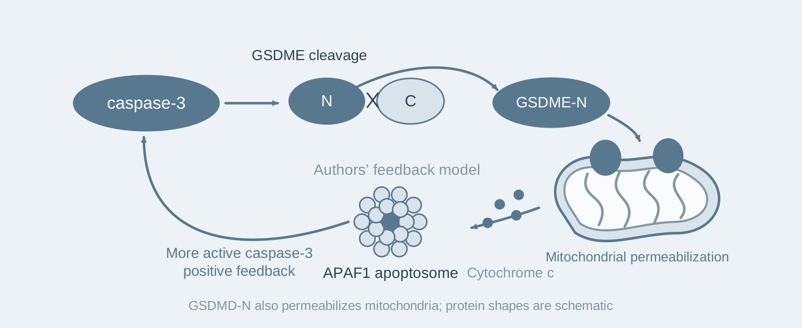
Key mechanism: GSDME-N permeabilizes the outer mitochondrial membrane to release cytochrome c, activating more caspase-3 through the apoptosome and creating a positive feedback loop;
- 03
Significance: Mitochondrial permeabilization is a shared property of the gasdermin family; GSDMD-N and GSDMA-N can also do this, allowing inflammasome activation to connect to the apoptotic pathway.
On this page
1. What question does this paper answer?
GSDME executes pyroptosis after cleavage by caspase-3, an established downstream function. This paper asks: Does GSDME also influence the apoptotic pathway itself in return? The answer is yes: besides forming pores in the plasma membrane, GSDME-N permeabilizes mitochondria to release cytochrome c and amplifies caspase-3 activation through the APAF1 apoptosome. GSDMD-N has the same capability.
2. Following the chain of evidence
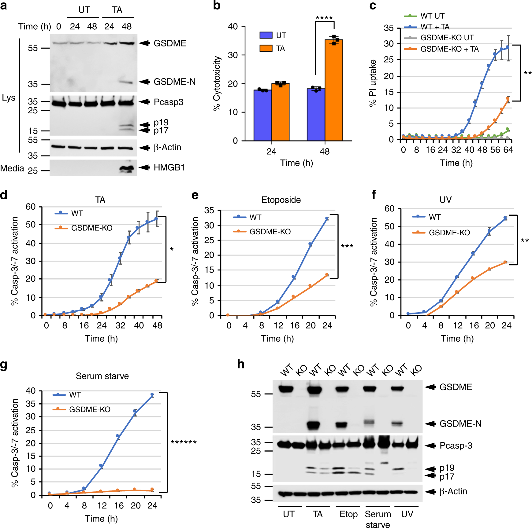
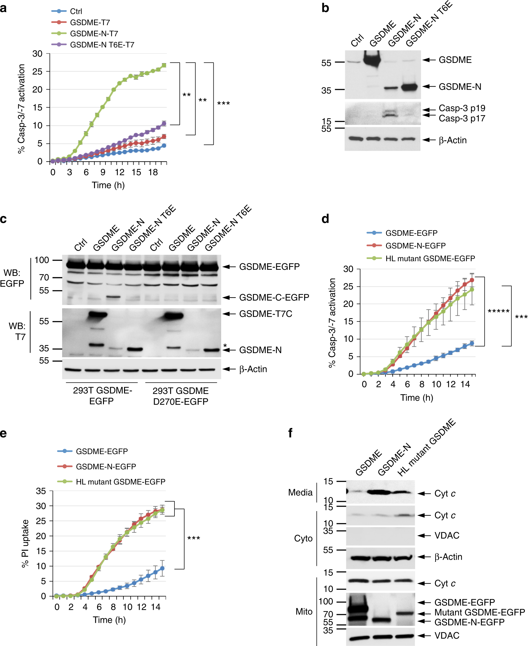
Step 1: Discover that GSDME influences caspase-3 activation (Figs. 1–3). The authors induced apoptosis with a glucocorticoid in CEM-C7 leukemia cells. After CRISPR deletion of GSDME, not only was pyroptosis reduced, but the speed and magnitude of caspase-3 activation also decreased markedly. This phenotype was reproducible in immortalized macrophages, melanoma cells, thymoma cells and with other apoptotic stimuli. The authors also found that a phosphomimetic mutation of threonine at position 6 of GSDME, T6E, disrupted oligomerization and thereby abolished pore-forming activity; caspase-3 activation was likewise weaker in cells reconstituted with the T6E mutant. These results show that GSDME's enhancement of caspase-3 depends on its pore-forming activity itself, rather than other functions of the cleaved fragment.
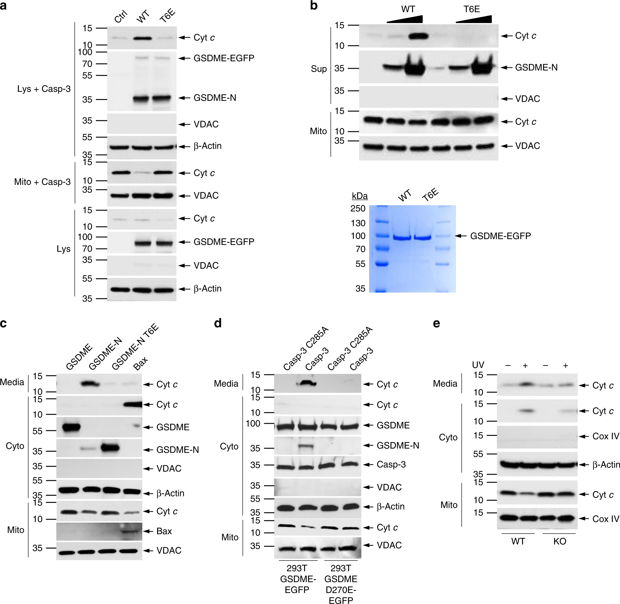
Step 2: Demonstrate that GSDME-N directly permeabilizes mitochondria (Figs. 4–5). Imaging showed that GSDME-N colocalized with mitochondria, and the mitochondrial protein HtrA2 was released before plasma membrane rupture. In vitro, the authors incubated purified mitochondria with caspase-3-cleaved GSDME and directly observed cytochrome c release, whereas the T6E mutant had no effect. These results show that GSDME-N can directly permeabilize the outer mitochondrial membrane, releasing cytochrome c and other pro-apoptotic factors.
Step 3: Confirm an amplification pathway operating in parallel with tBid (Figs. 6–7). In death receptor signaling, caspase-8 activates the mitochondrial pathway by cleaving Bid. The authors tested whether GSDME was redundant with this mechanism. In TNFα-treated CEM-C7 cells, deleting either Bid or GSDME slowed caspase-3 activation, and deleting both weakened it further. In APAF1-knockout cells, ectopically expressed GSDME-N could not activate caspase-3 at all, showing that this amplification loop must act through the apoptosome. These results show that GSDME-N amplifies caspase-3 activation through the mitochondria–apoptosome pathway, resulting in cleavage of more GSDME and creating a positive feedback loop that operates in parallel with the tBid pathway.
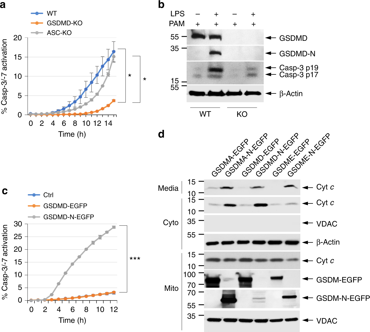
Step 4: Extend the findings to GSDMD and in vivo (Figs. 8–9). In macrophages transfected with cytosolic LPS, the authors found that GSDMD deletion markedly delayed and weakened caspase-3 activation, whereas ASC deletion had no effect. Overexpression of either GSDMD-N or GSDMA-N also released cytochrome c and activated caspase-3. In vivo, GSDME-knockout B16 melanomas grew faster after subcutaneous implantation in mice. These results show that mitochondrial permeabilization is a shared property of the gasdermin family and that GSDME's pro-death function has tumor-suppressive significance.
3. Implications for experiments
- Changes in apoptotic markers after gasdermin deletion should not be surprising: GSDME/GSDMD deletion can slow caspase-3 activation itself, a factor to consider when interpreting caspase-3 cleavage bands.
- Distinguish the timing of mitochondrial release from plasma membrane rupture: Mitochondrial proteins are released before plasma membrane rupture; readouts that conflate the two into a single time point would miss the direct action of GSDME-N.
- Pay attention to phosphorylation-dependent regulation: The T6E phosphomimetic mutation allows GSDME-N to reach the membrane but prevents oligomerization. Detecting "membrane localization" is not the same as detecting "activity".
4. Connections with other papers on this site
- Extension | GSDME mediates secondary necrosis following apoptosis (Rogers 2017, Nat Commun): The 2017 paper showed that GSDME determines whether apoptotic cells undergo membrane rupture; this paper shows that GSDME also amplifies upstream caspase-3 activation. Together, the two papers provide a complete picture of GSDME function.
- Comparison | Caspase-1 initiates apoptosis in the absence of GSDMD (Tsuchiya 2019, Nat Commun): Tsuchiya's paper reports that, in the absence of GSDMD, caspase-1 connects to mitochondrial apoptosis through Bid. This paper reports direct mitochondrial permeabilization by GSDMD-N. Both routes lead to the mitochondria.
- Extension | GSDMD permeabilizes mitochondria to amplify pyroptosis (Miao 2023, Immunity): This paper shows that gasdermin-N permeabilizes mitochondria to amplify caspase-3 during apoptosis. Miao shows that GSDMD-NT permeabilizes both mitochondrial membranes and amplifies pyroptosis itself in a cardiolipin-dependent manner—the mechanism of "gasdermin attacking mitochondria" serves two cell death pathways.
- Extension | Effector caspases amplify apoptotic signaling through feedback (McComb 2019, Sci Adv): Positive feedback in apoptotic signaling operates at two levels. McComb shows that effector caspases amplify upstream caspase-8/-9 and mitochondrial permeabilization through protein cleavage; this paper shows that GSDME-N amplifies caspase-3 activation itself by permeabilizing mitochondria.
(The cross-paper comparisons above are a synthesis, rather than direct conclusions of any one original paper.)
What remains unresolved
Limitations: ① The specific molecular mechanism by which GSDME-N permeabilizes mitochondria, including whether it binds cardiolipin, was not established; ② the upstream kinase and physiological setting of T6 phosphorylation were not determined.
Gasdermin pores permeabilize mitochondria to augment caspase-3 activation during apoptosis and inflammasome activation
Corey Rogers; Dan A Erkes; Alexandria Nardone; Andrew E Aplin; Teresa Fernandes-Alnemri; Emad S Alnemri Nature Communications. 2019;10:.
Figures are from the verified original paper stored in the library. Original labels, axes and controls are retained; figure numbers and sources are registered in the library.
References
- Rogers C, Erkes DA, Nardone A, et al. Gasdermin pores permeabilize mitochondria to augment caspase-3 activation during apoptosis and inflammasome activation. Nat Commun. 2019;10:1689. doi:10.1038/s41467-019-09397-2
- Rogers C, Fernandes-Alnemri T, Mayes L, et al. Cleavage of DFNA5 by caspase-3 during apoptosis mediates progression to secondary necrotic/pyroptotic cell death. Nat Commun. 2017;8:14128.
- Tsuchiya K, Nakajima S, Hosojima S, et al. Caspase-1 initiates apoptosis in the absence of gasdermin D. Nat Commun. 2019;10:2091.
Paper discussion
No sign-up. Nickname optional.