- 01
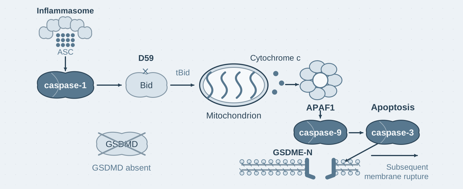
Main finding: In the absence of GSDMD, caspase-1 activation initiates caspase-3-dependent apoptosis rather than pyroptosis; caspase-3 then cleaves GSDME to drive subsequent membrane rupture;
- 02
Key mechanism: Caspase-1 cleaves Bid at D59, activating caspase-3 through the tBid–cytochrome c–apoptosome–caspase-9 axis;
- 03
Significance: The level of GSDMD expression determines the mode of death downstream of caspase-1. In cells with naturally low GSDMD expression, such as cortical neurons, inflammasome activation can manifest as apoptosis.
On this page
1. What question does this paper answer?
The accepted outcome after inflammasomes activate caspase-1 is GSDMD-mediated pyroptosis. This paper asks: What does caspase-1 activation trigger in cells without GSDMD? Its conclusion is that caspase-1 cleaves Bid to initiate the mitochondrial apoptotic pathway. The cells die by apoptosis, after which caspase-3 cleaves GSDME to drive membrane rupture.
2. Following the chain of evidence
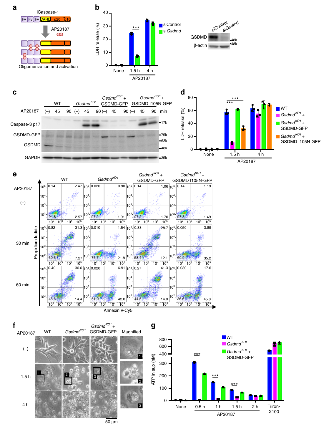
Step 1: Establish that death in the absence of GSDMD depends on caspase-1 and has apoptotic features (Figs. 1–2). The authors transfected Gsdmd−/− macrophages with poly(dA:dT) or infected them with Salmonella. LDH release was slower than in wild-type cells but remained substantial. The cells showed apoptotic morphology, including membrane blebbing, and caspase-3, -8, and -9 were all cleaved and activated. Casp1−/− cells did not show this response. ASC-deficient RAW264.7 cells behaved similarly, ruling out the ASC–caspase-8 pathway. To test the capacity of caspase-1 directly, the authors used a drug-induced caspase-1 dimerization system in colon cancer cells. After Gsdmd knockout, caspase-1 activation caused apoptosis. Re-expression of wild-type GSDMD restored pyroptosis, whereas re-expression of the pore-defective I125N mutant did not. Together, these results indicate that whether cells undergo pyroptosis or apoptosis downstream of caspase-1 is determined by the pore-forming activity of GSDMD.
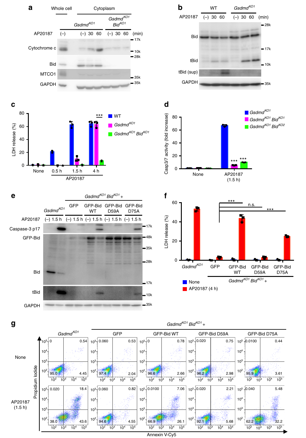
Step 2: Define the apoptotic pathway (Figs. 3–6). The authors examined downstream molecules one by one in Gsdmd-knockout cells. Caspase-3 knockout almost completely blocked death, and caspase-9 knockout markedly reduced caspase-3 activation, whereas knocking out caspase-2, -6, -7, or -8 had no effect. Caspase-1 was already known to cleave Bid in vitro. The authors found that after caspase-1 activation, Bid was cleaved to tBid, accompanied by mitochondrial depolarization, cytochrome c release, and formation of an APAF1–caspase-9 complex. Bid knockout severely delayed these events. Re-expression experiments identified the cleavage site: wild-type Bid or the D75A mutant restored apoptosis, whereas D59A did not. Together, these results indicate that caspase-1 cleaves Bid at D59 and activates caspase-3 through the mitochondrial–caspase-9 axis.
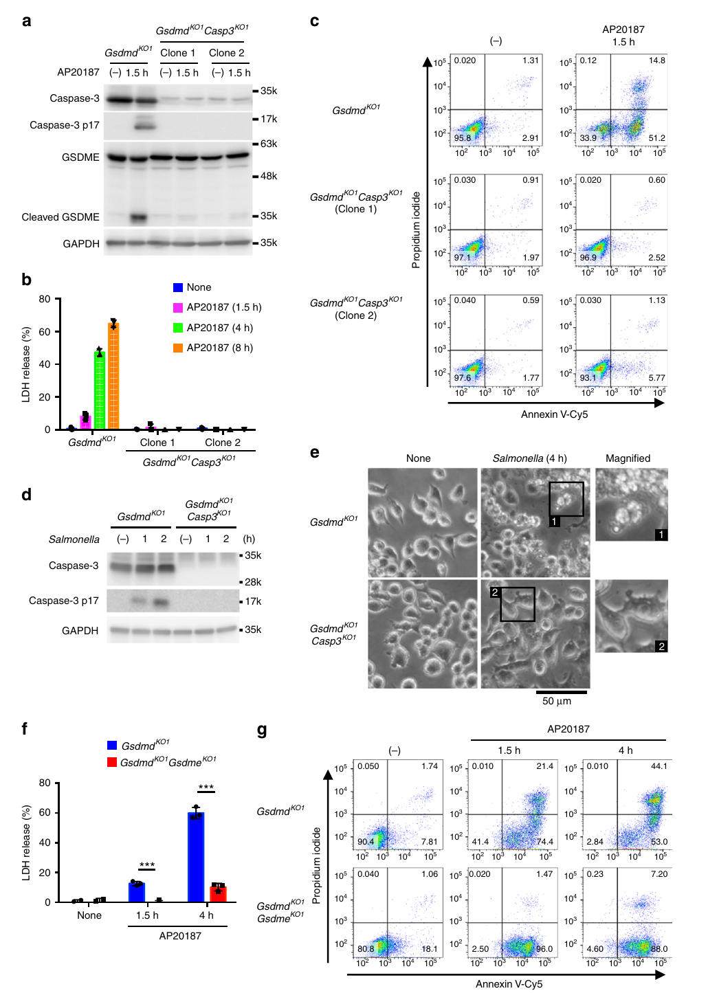
Step 3: Answer "What releases the LDH?" (Fig. 3). What executes LDH release in GSDMD-deficient cells? The authors found that activated caspase-3 cleaves GSDME. GSDME knockout substantially reduced early membrane damage, measured by PI uptake and LDH release, but did not affect phosphatidylserine (PS) externalization or membrane blebbing. Together, these results indicate that apoptosis initiated by caspase-1 rapidly progresses to secondary necrosis through caspase-3–GSDME.
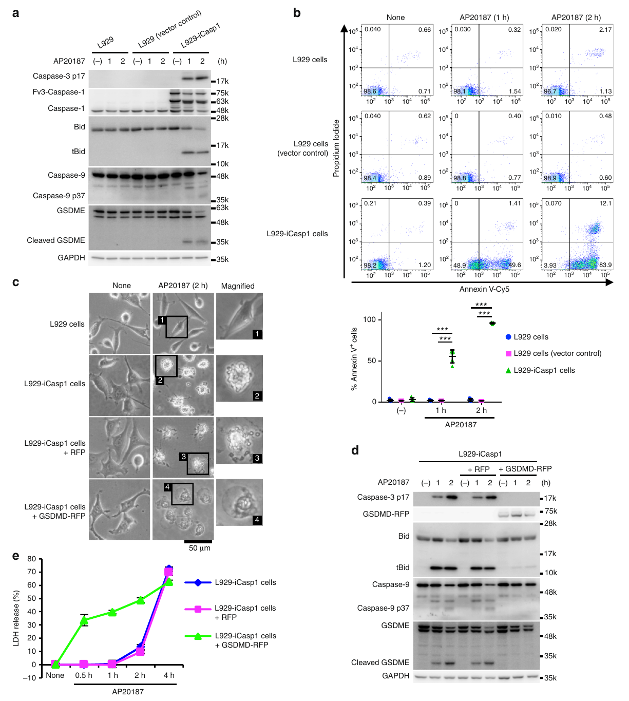
Step 4: Test physiological relevance (Fig. 9). The authors examined L929 cells and mouse cortical neurons, which naturally do not express GSDMD, and found that caspase-1 activation likewise induced Bid-dependent apoptosis. In Salmonella infection experiments, intracellular bacterial survival increased in Bid-knockout cells. Together, these results indicate that this apoptotic pathway exists in naturally GSDMD-negative cells and can substitute for pyroptosis to restrict intracellular bacteria.
3. Implications for experiments
- LDH release from Gsdmd-knockout cells does not necessarily mean pyroptosis: In this paper, it arose from secondary necrosis after apoptosis. Caspase-3 and GSDME cleavage, together with cell morphology, are needed to determine the mode of death.
- Check basal expression in inflammasome experiments using Gsdmd-knockout cells: In cells with naturally low GSDMD expression, such as L929 cells and neurons, caspase-1 activation already leads to apoptosis.
- Bid connects caspase-1 to the apoptotic pathway: Detecting tBid and cleavage at D59 provides the most direct readout of whether this pathway is involved.
4. Connections to other papers on this site
- Extension | GSDMD pyroptosis (He 2015, Cell Res): He et al. established caspase-1–GSDMD as the canonical pyroptotic axis. This paper describes the alternative route taken by caspase-1 when GSDMD is absent. Together, the two papers provide a complete branching map downstream of caspase-1.
- Extension | GSDME-mediated secondary necrosis after apoptosis (Rogers 2017, Nat Commun): Secondary necrosis after apoptosis in this paper is executed by caspase-3–GSDME, consistent with the mechanism described by Rogers et al.
- Comparison | Gasdermins permeabilize mitochondria to amplify caspase-3 (Rogers 2019, Nat Commun): In this paper, caspase-1 connects indirectly to mitochondria through Bid. In Rogers et al., gasdermin-N directly permeabilizes mitochondria. Published in the same journal in the same year, these two routes complement one another.
- Extension | Caspase-1 self-cleavage terminates inflammasome activity (Boucher 2018, JEM): Boucher et al. describe an approximately 30-minute window of caspase-1 activity. Bid cleavage and the initiation of apoptosis in this paper occur within this window.
(The comparisons above are integrated interpretations across papers, rather than direct conclusions of any single original paper.)
What remains unresolved
Limitations: ① A second, Bid-independent apoptotic pathway was observed only in certain cell lines, and its mechanism remains unclear; ② The role of GSDME in primary macrophages differs between this paper and other reports, requiring validation in double-knockout mice.
Caspase-1 initiates apoptosis in the absence of gasdermin D
Tsuchiya, Kohsuke; Nakajima, Shinsuke; Hosojima, Shoko; Thi Nguyen, Dinh; Hattori, Tsuyoshi; Manh Le, Thuong; Hori, Osamu; Mahib, Mamunur Rashid; Yamaguchi, Yoshifumi; Miura, Masayuki; Kinoshita, Takeshi; Kushiyama, Hiroko; Sakurai, Mayumi; Shiroishi, Toshihiko; Suda, Takashi Nature Communications. 2019;10:.
Figures are from the verified original paper stored in the library. Original labels, axes and controls are retained; figure numbers and sources are registered in the library.
References
- Tsuchiya K, Nakajima S, Hosojima S, et al. Caspase-1 initiates apoptosis in the absence of gasdermin D. Nat Commun. 2019;10:2091. doi:10.1038/s41467-019-09753-2
- He WT, Wan H, Hu L, et al. Gasdermin D is an executor of pyroptosis and required for interleukin-1β secretion. Cell Res. 2015;25(12):1285-1298.
- Rogers C, Fernandes-Alnemri T, Mayes L, et al. Cleavage of DFNA5 by caspase-3 during apoptosis mediates progression to secondary necrotic/pyroptotic cell death. Nat Commun. 2017;8:14128.
Paper discussion
No sign-up. Nickname optional.