- 01
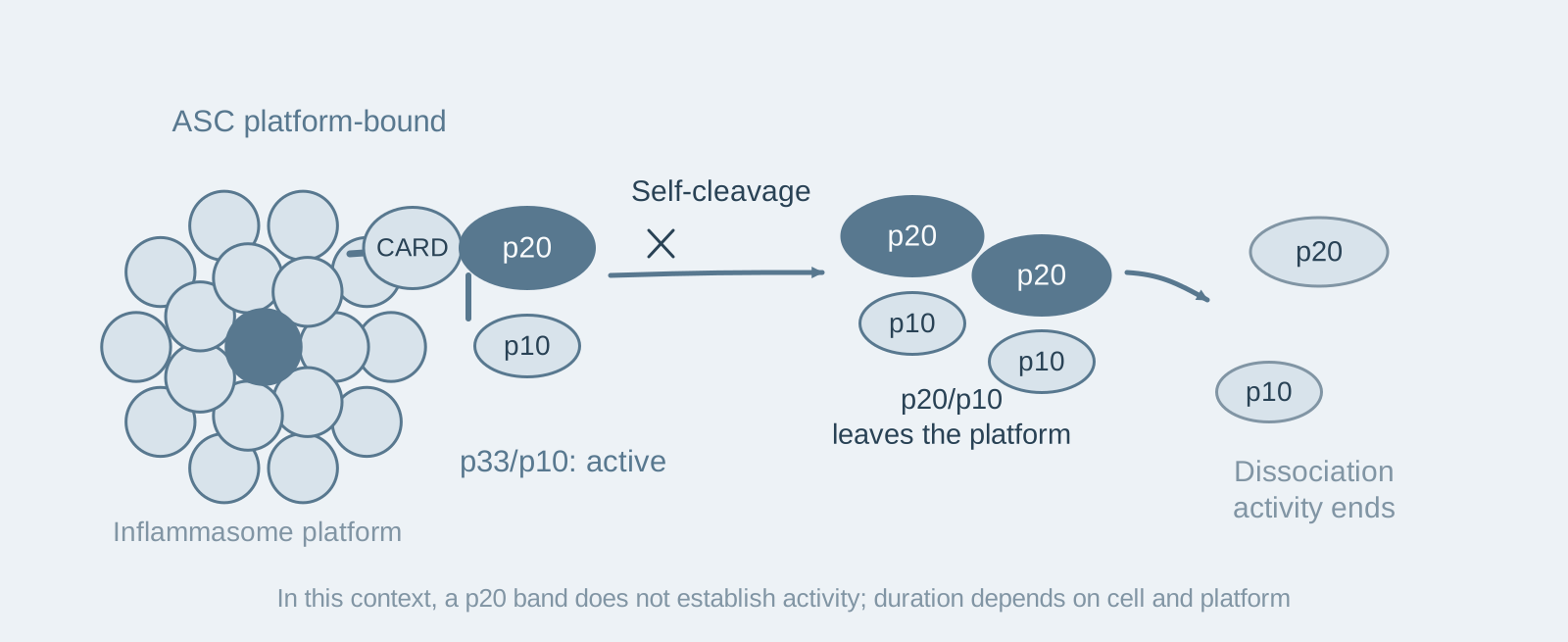
Main finding: The principal active form of caspase-1 in cells is p33/p10; activity disappears within about 30 minutes after nigericin stimulation, and p20/p10 is an inactive product;
- 02
Key mechanism: Self-cleavage of p33/p10 at the linker produces p20/p10, releasing it from the inflammasome complex, after which tetramer dissociation inactivates it;
- 03
Significance: The inflammasome–caspase-1 complex is a holoenzyme with an intrinsic time limit, and its activity duration is determined by inflammasome platform size and cell type.
On this page
I. What question does this paper answer?
Direct evidence had been lacking on how long caspase-1 activity lasts after inflammasome activation and how it is shut down. This paper asks: Which form of caspase-1 is actually active in cells, and how is its activity terminated? Its conclusion is that the active form is inflammasome-bound p33/p10, which becomes inactive by self-cleaving to produce p20/p10 and leaving the complex; the entire process takes about 30 minutes.
II. Following the logic of the evidence
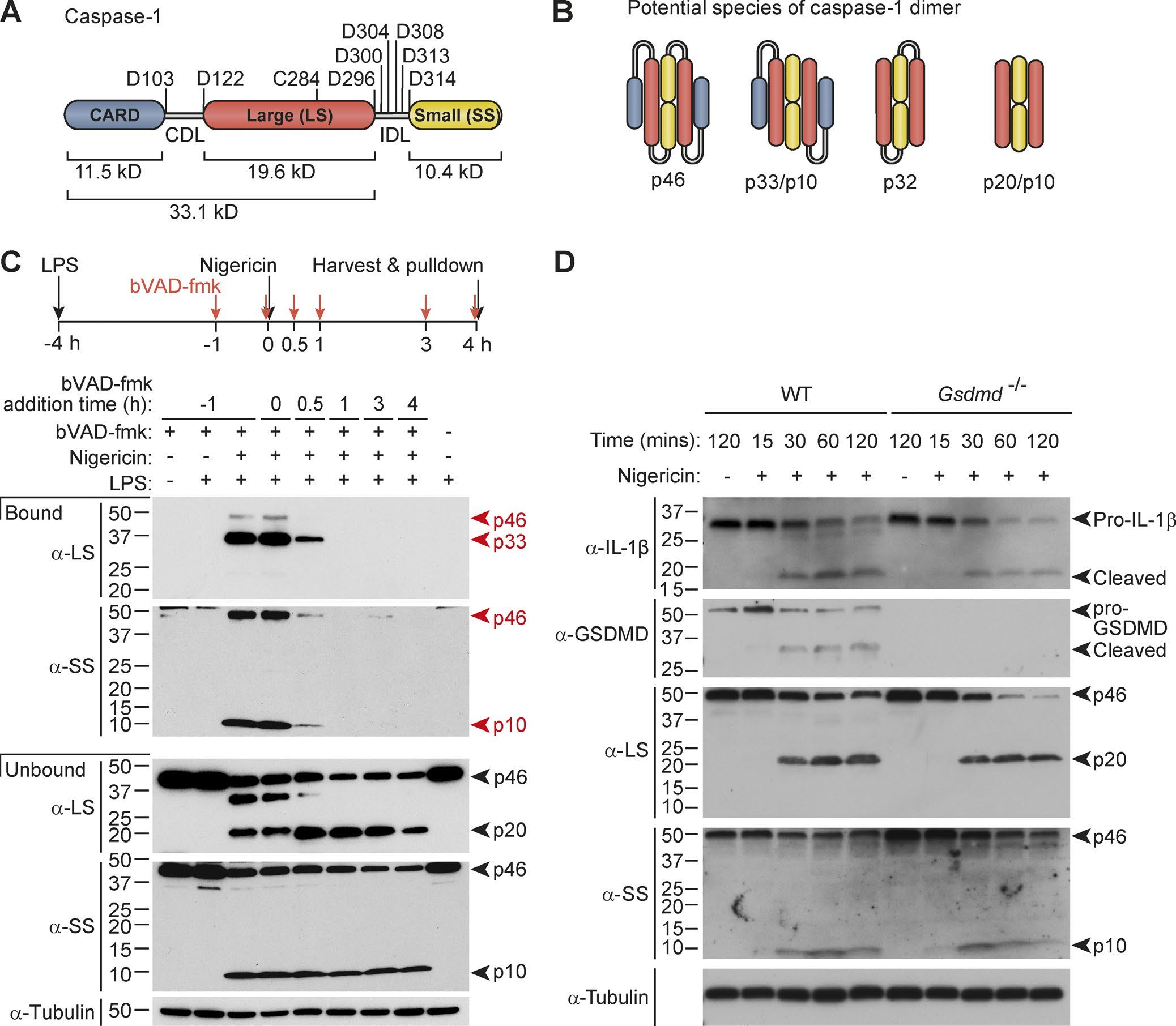
Step 1: Identify the active form in cells (Figure 1). In LPS-primed macrophages stimulated with nigericin, the authors used bVAD-fmk, a probe that covalently binds the active site, to capture active caspase-1 at different time points. The main species pulled down was p33/p10, with almost no p20; adding the probe after 30 minutes of stimulation captured nothing. Substrate cleavage followed the same timing: cleavage of pro-IL-1β and pro-GSDMD was concentrated at 15–30 minutes after stimulation; delaying cell lysis through Gsdmd deletion or glycine did not extend this window. Together, these results show that the active caspase-1 species in cells is p33/p10 rather than p20/p10, the activity window is about 30 minutes, and its closure is independent of cell lysis.
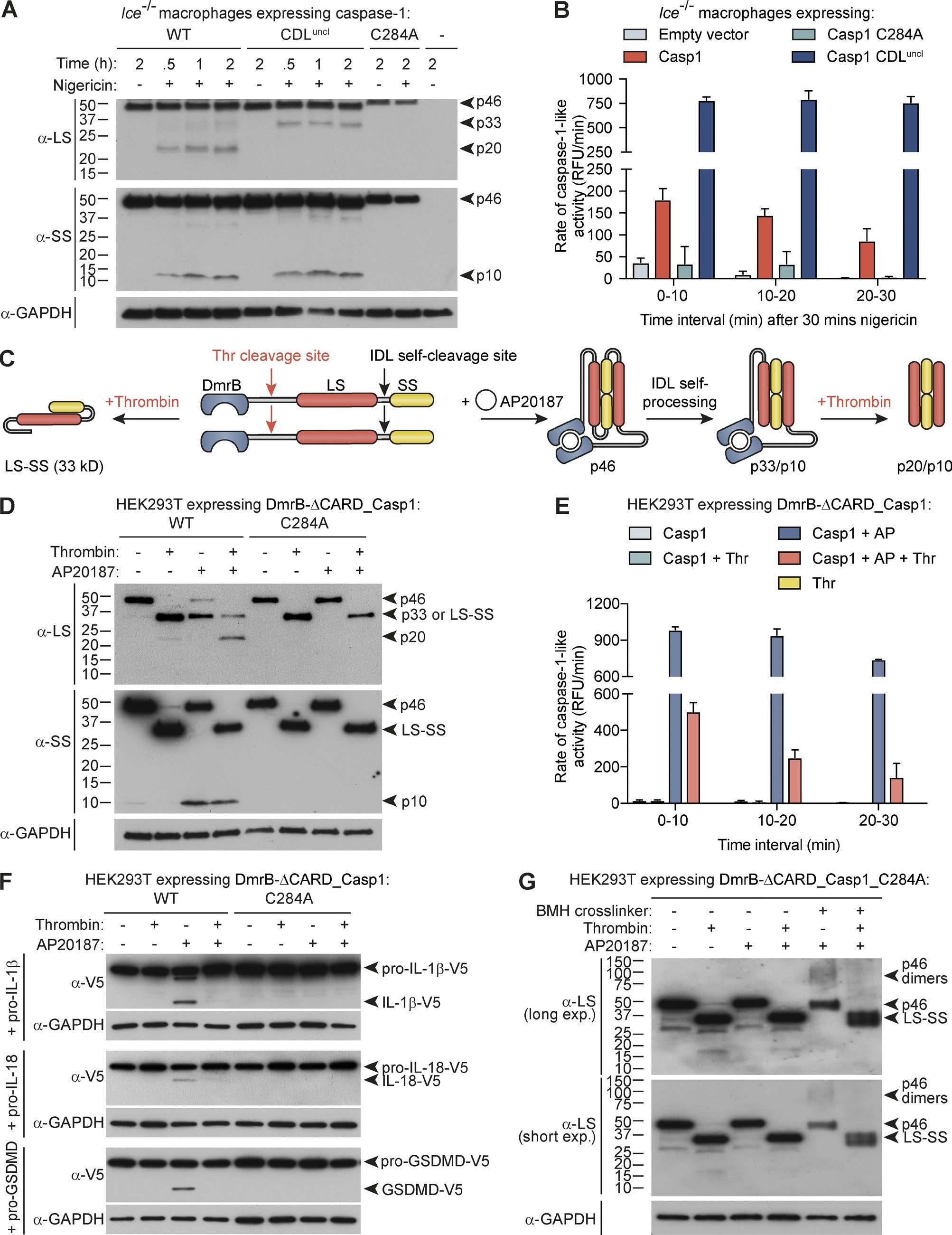
Step 2: Demonstrate that self-cleavage causes inactivation (Figure 2). The authors reconstituted knockout cells with a caspase-1 mutant whose linker could not be cleaved, and found stronger, longer-lasting activity than with wild-type caspase-1. Conversely, when they used thrombin to cleave the linker of an inducibly dimerized caspase-1, the dimer dissociated and activity rapidly disappeared, together with its ability to cleave pro-GSDMD and pro-IL-1β. Recombinant p20/p10 was active at high concentrations but became inactive through tetramer dissociation when diluted to concentrations near those in cells, explaining why in vitro biochemical experiments and cellular experiments reached opposite conclusions. Together, these results show that self-cleavage at the linker releases p20/p10 from the inflammasome, followed by tetramer dissociation and inactivation—this is an intrinsic mechanism for terminating caspase-1 activity.
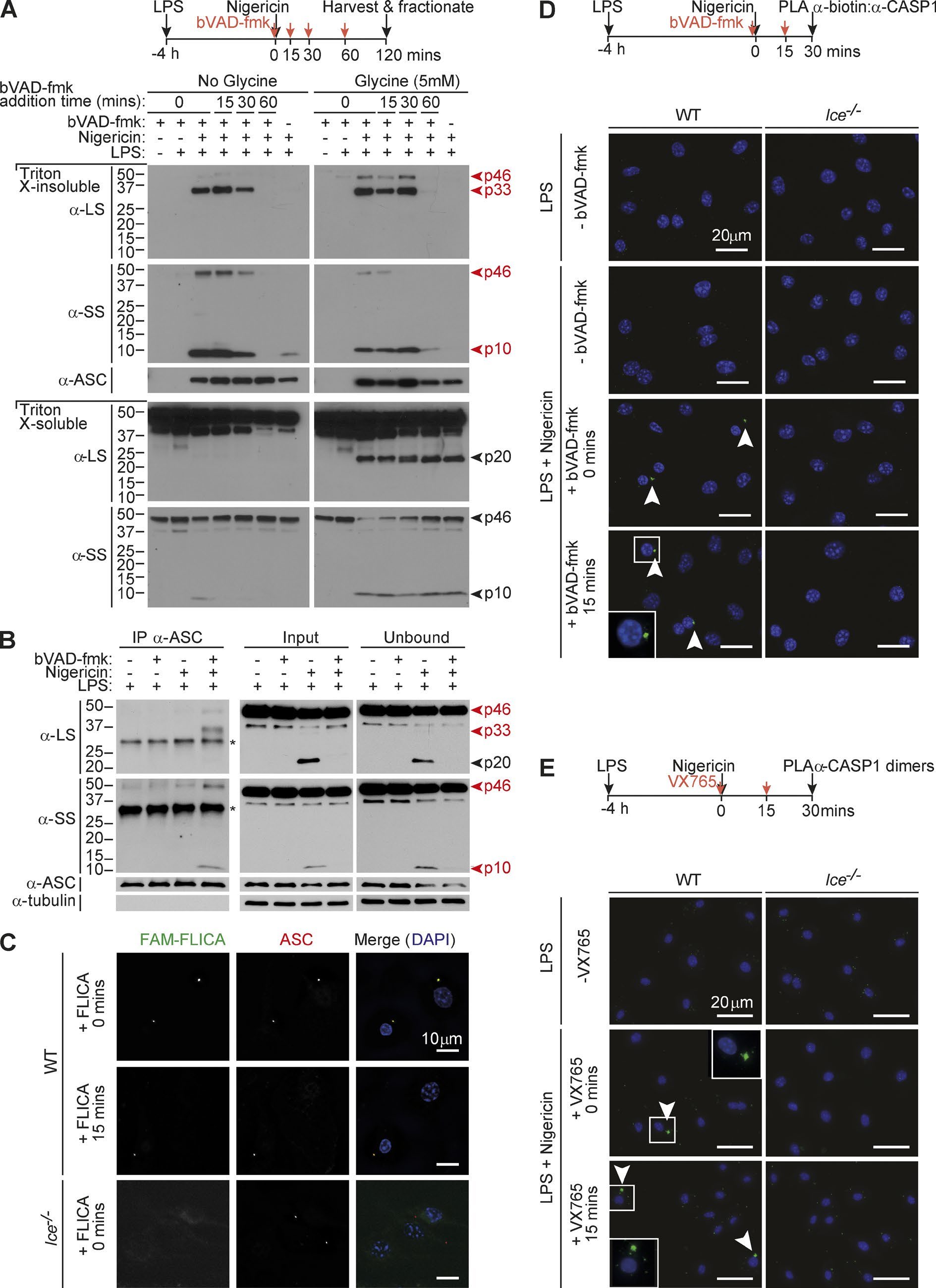
Step 3: Locate where activity occurs (Figure 3). The authors isolated the ASC speck fraction (the speck is the inflammasome complex formed by ASC polymerization) and performed co-immunoprecipitation and proximity ligation assays. They found p33/p10 bound to ASC, whereas p20 did not co-immunoprecipitate with ASC; both the activity-probe signal and dimerized caspase-1 localized to the ASC speck. Together, these results show that caspase-1 activity occurs on the inflammasome complex and is lost after release.
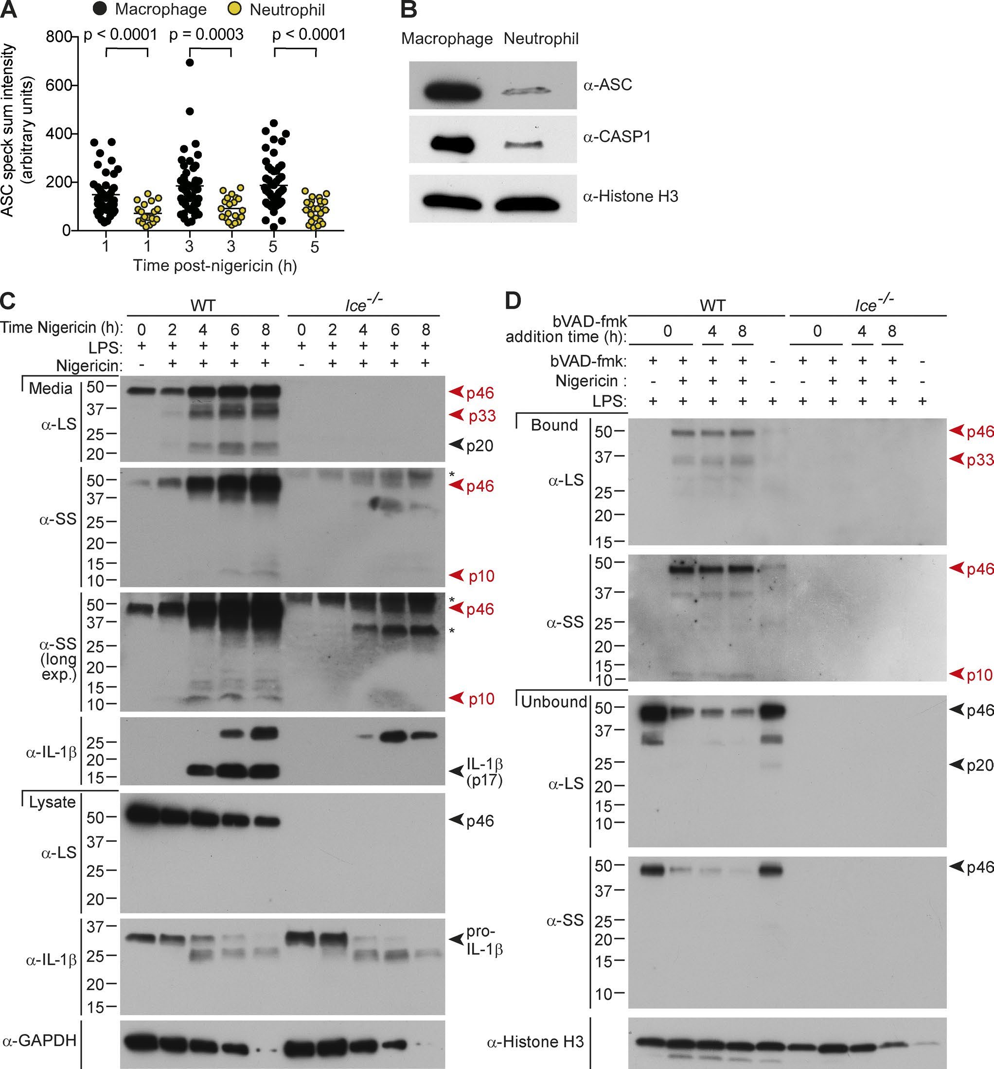
Step 4: Test what determines activity duration (Figures 4–5). When the authors activated NLRC4 with flagellin in Asc−/− macrophages, caspase-1 was predominantly in its full-length p46 form and activity persisted longer; in Asc+/− cells, specks were half the size and activity duration lay between the two. Consistent with this, in neutrophils with lower ASC expression and smaller specks, caspase-1 activity could persist for 8 hours, and IL-1β secretion continued to increase up to 6 hours. Together, these results show that the size of the inflammasome platform determines the efficiency of caspase-1 self-cleavage and thus its activity duration; this is one reason why the timing of inflammasome responses differs between macrophages and neutrophils.
III. Implications for experiments
- The p20 band on a Western blot cannot be treated as an activity indicator: p20 is a product left after inactivation, and more p20 actually indicates that the activity window has closed; measuring activity requires activity probes such as FLICA or bVAD.
- Consider ASC expression when comparing inflammasome responses across cell types: Cells with less ASC and smaller specks have longer-lasting caspase-1 activity; this is a confounding factor when comparing IL-1β secretion kinetics.
- The cytokine-processing window is short: In macrophages, IL-1β processing is concentrated at 15–30 minutes after stimulation, so the sampling schedule should cover this window.
IV. Connections to other papers on this site
- Extension | Caspase-1 initiates apoptosis through Bid (Tsuchiya 2019, Nat Commun): In GSDMD-deficient cells, caspase-1 turns to cleaving Bid, also within the approximately 30-minute activity window defined in this paper.
- Comparison | RIPK3 cleavage limits NLRP3 (Tran 2024, Cell Death Differ): This paper describes caspase-1 self-cleavage limiting the duration of inflammasome activity, whereas Tran describes caspase-8 cleavage of RIPK3 limiting the intensity of inflammasome activation; each pro-inflammatory pathway has its own negative-feedback mechanism.
- Extension | The caspase-1 inflammasome activates a caspase-7 cascade (Lamkanfi 2008, Mol Cell Proteomics): Caspase-1 cleavage events have two functional outcomes—Lamkanfi demonstrates that caspase-1 cleaves and activates caspase-7 (signal transmission), whereas this paper demonstrates that caspase-1 self-cleavage releases it from the inflammasome and inactivates it (signal termination).
(The comparisons across papers above are a synthesis, rather than a direct conclusion of any one original paper.)
What remains unresolved
Limitations: ① Binding of the activity probe bVAD-fmk to the active site itself stabilizes the captured form and may slightly interfere with kinetic measurements. ② The mechanism was established mainly in mouse macrophages and neutrophils.
Caspase-1 self-cleavage is an intrinsic mechanism to terminate inflammasome activity
Dave Boucher; Mercedes Monteleone; Rebecca C. Coll; Kaiwen W. Chen; Connie M. Ross; Jessica L. Teo; Guillermo A. Gomez; Caroline L. Holley; Damien Bierschenk; Katryn J. Stacey; Alpha S. Yap; Jelena S. Bezbradica; Kate Schroder Journal of Experimental Medicine. 2018;215:827–840.
Figures are from the verified original paper stored in the library. Original labels, axes and controls are retained; figure numbers and sources are registered in the library.
References
- Boucher D, Monteleone M, Coll RC, et al. Caspase-1 self-cleavage is an intrinsic mechanism to terminate inflammasome activity. J Exp Med. 2018;215(3):827-840. doi:10.1084/jem.20172222
- Broz P, von Moltke J, Jones JW, et al. Differential requirement for caspase-1 autoproteolysis in pathogen-induced cell death and cytokine processing. Cell Host Microbe. 2010;8(6):471-483.
- Chen KW, Groß CJ, Sotomayor FV, et al. The neutrophil NLRC4 inflammasome selectively promotes IL-1β maturation without pyroptosis during acute Salmonella challenge. Cell Rep. 2014;8(2):570-582.
Paper discussion
No sign-up. Nickname optional.