- 01
Main finding: Blocking caspase-8-mediated RIPK3 cleavage (D333A knock-in) does not cause uncontrolled necroptosis; the mice are healthy and fertile;
- 02
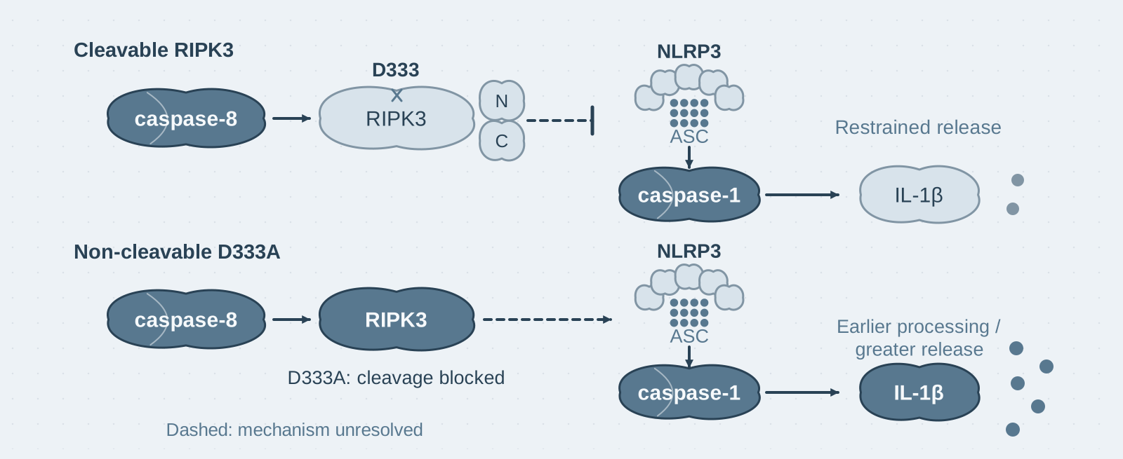
Key mechanism: When IAPs are inhibited, RIPK3 cleavage acts as negative feedback, restricting TLR–RIPK3-driven NLRP3 activation and IL-1β secretion;
- 03
Significance: Revises the textbook model and explains the high conservation of this site—it regulates the intensity of inflammation, rather than determining whether a cell lives or dies.
On this page
I. What this paper answers
Caspase-8 activity is a key factor limiting necroptosis: when it is knocked out, mice die during embryogenesis because RIPK3–MLKL activity becomes uncontrolled. The textbook model therefore holds that caspase-8 must cleave a substrate to inhibit necroptosis. However, knock-in mouse studies in recent years have ruled out candidate substrates one by one: cleavage of CYLD and cFLIP is not required; RIPK1 cleavage is indeed required, but its role is to prevent apoptosis and autoinflammation, rather than necroptosis. RIPK3 became the last candidate—caspase-8 cleaves it at D333, and this site is strictly conserved across 94 vertebrate species. If this cleavage is truly critical for preventing death, mutating the site should reproduce the lethal phenotype.
II. The logic of the evidence
The argument has two parts: first test the "prevention of necroptosis" hypothesis, then identify the actual function.
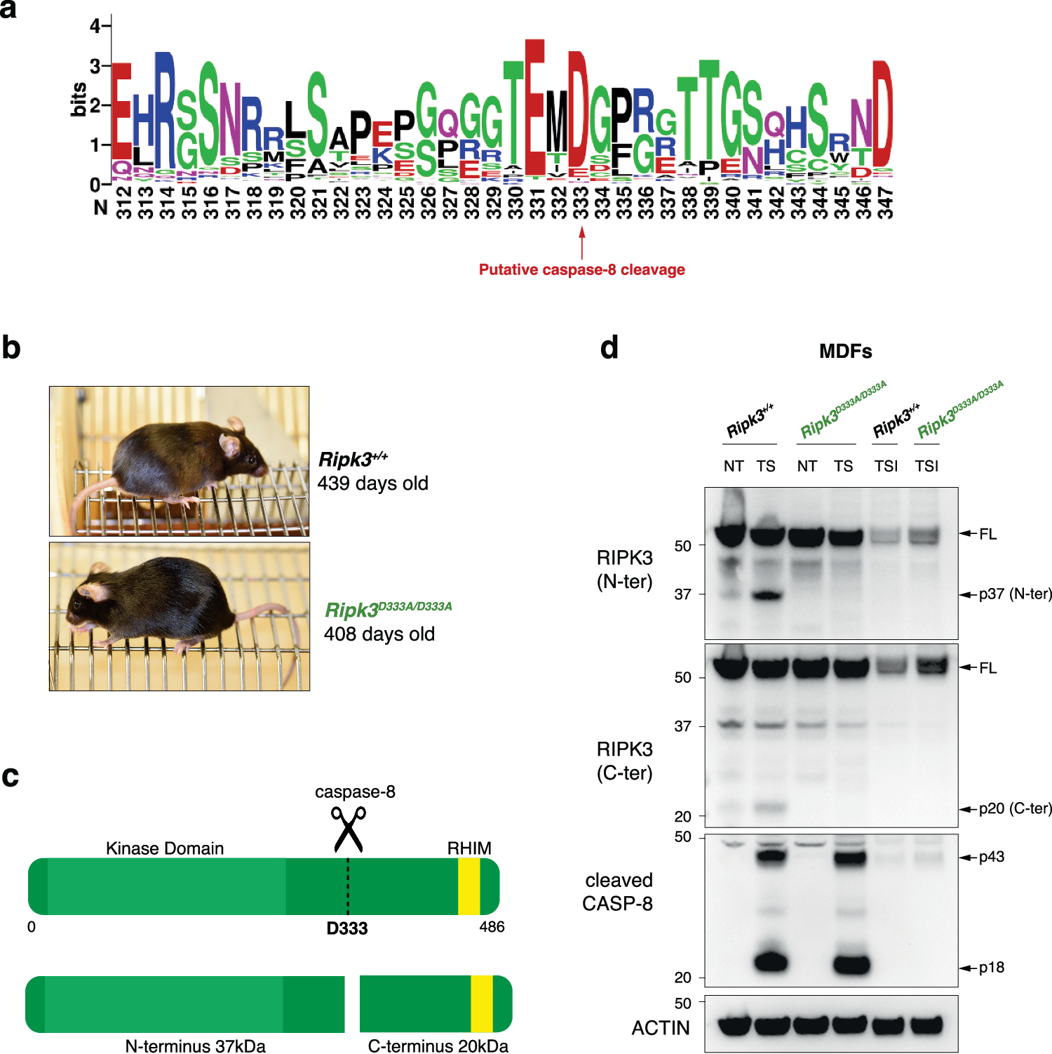
Step 1: Establish that cleavage actually occurs (Figure 1). The authors treated wild-type cells with TNF plus a Smac-mimetic and detected RIPK3 cleavage fragments of the expected sizes; these were not detected in mutant cells or when a caspase inhibitor was added. These results indicate that D333 is a genuine and unique cleavage site in vivo—without first establishing that cleavage really occurs, the subsequent "absence of a phenotype" provides no meaningful information.
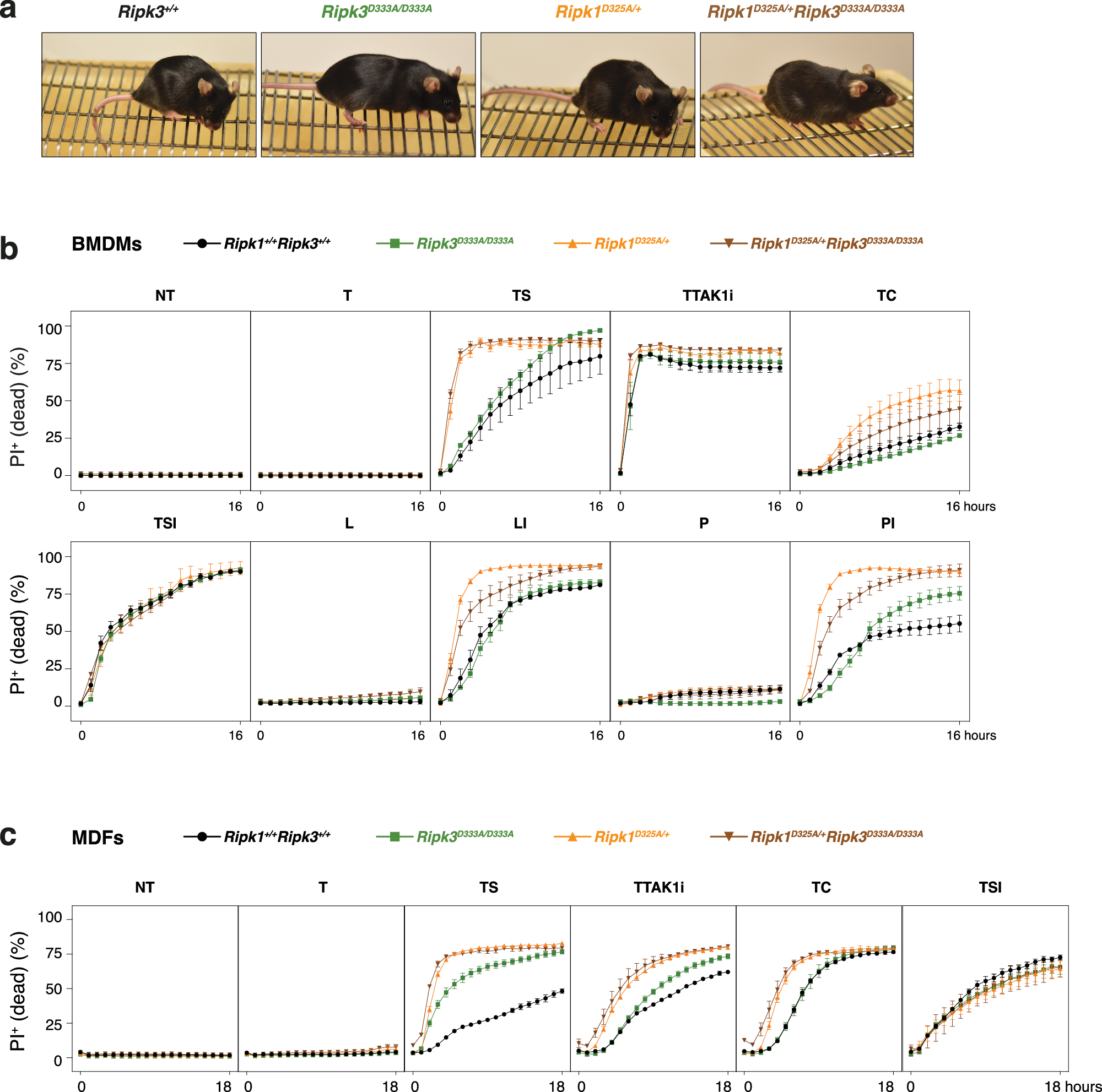
Step 2: Test the lethal phenotype at four levels (Figures 1–4). The authors observed that D333A knock-in mice were born at the expected Mendelian frequency and remained healthy for more than 400 days; when mutant cells were treated with combinations including TNF or LPS, their death kinetics and RIPK3/MLKL phosphorylation did not differ from those of wild-type cells; double-mutant mice obtained by crossing them with mice carrying uncleavable RIPK1 were also healthy, ruling out the alternative explanation that "RIPK1 cleavage compensates for the loss of RIPK3 cleavage". These results show that RIPK3 cleavage is not required to inhibit necroptosis.
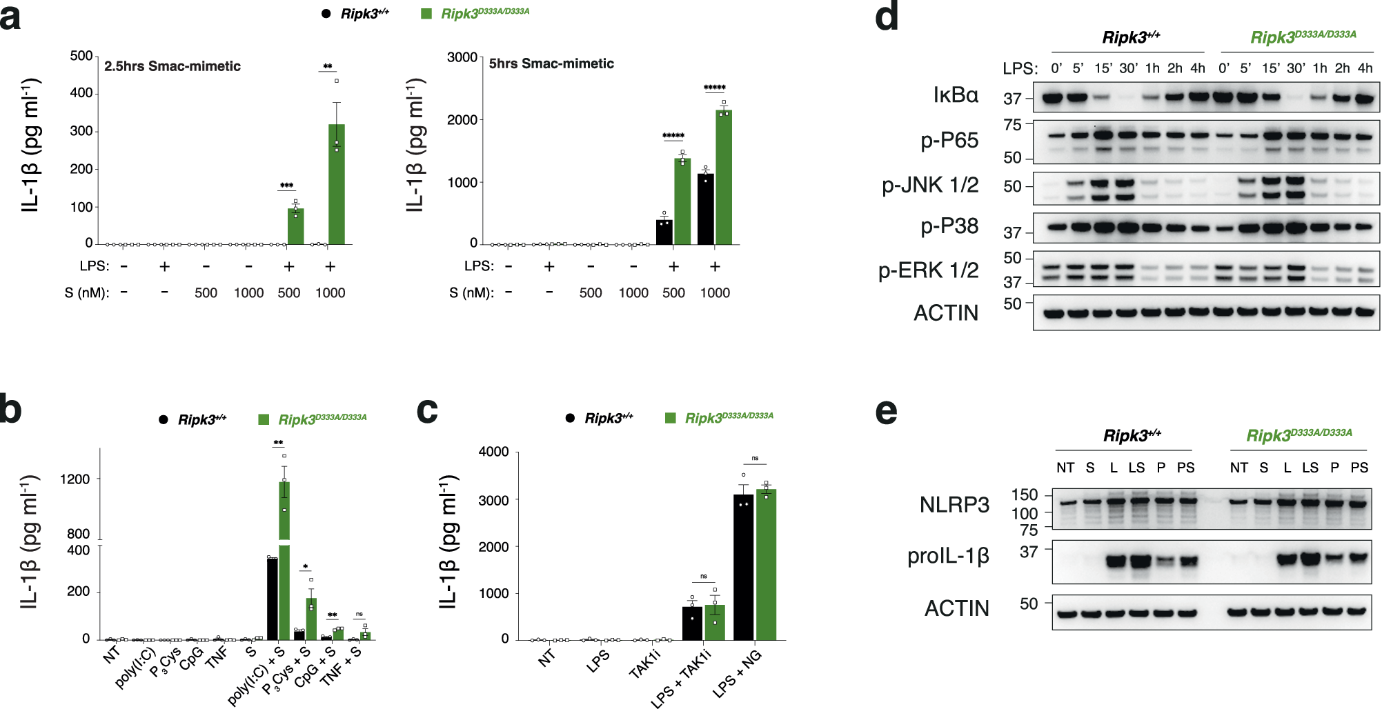
Step 3: Turn to inflammatory readouts (Figure 5). The authors primed macrophages with LPS and then added a Smac-mimetic, finding significantly higher IL-1β secretion in mutant cells than in wild-type cells; priming with other TLR ligands gave the same conclusion. Control experiments defined the scope of the effect: activators that do not act through RIPK3 (nigericin and a TAK1 inhibitor) were unaffected by the mutation; NF-κB and pro-IL-1β levels did not differ, ruling out an effect on priming (the preparatory step before inflammasome activation).
Step 4: Mechanistic and in vivo validation (Figure 6). Examining mutant cells, the authors found that cleavage of caspase-1, GSDMD and IL-1β appeared earlier than in wild-type cells, and the NLRP3 inhibitor MCC950 could reverse this phenomenon; following an in vivo LPS or poly(I:C) challenge, serum IL-1β was significantly higher in mutant mice than in wild-type mice. These results indicate that the actual function of RIPK3 cleavage is to provide negative feedback that restricts the TLR–RIPK3–NLRP3 axis; the substrate on which caspase-8 depends to inhibit necroptosis remains unidentified.
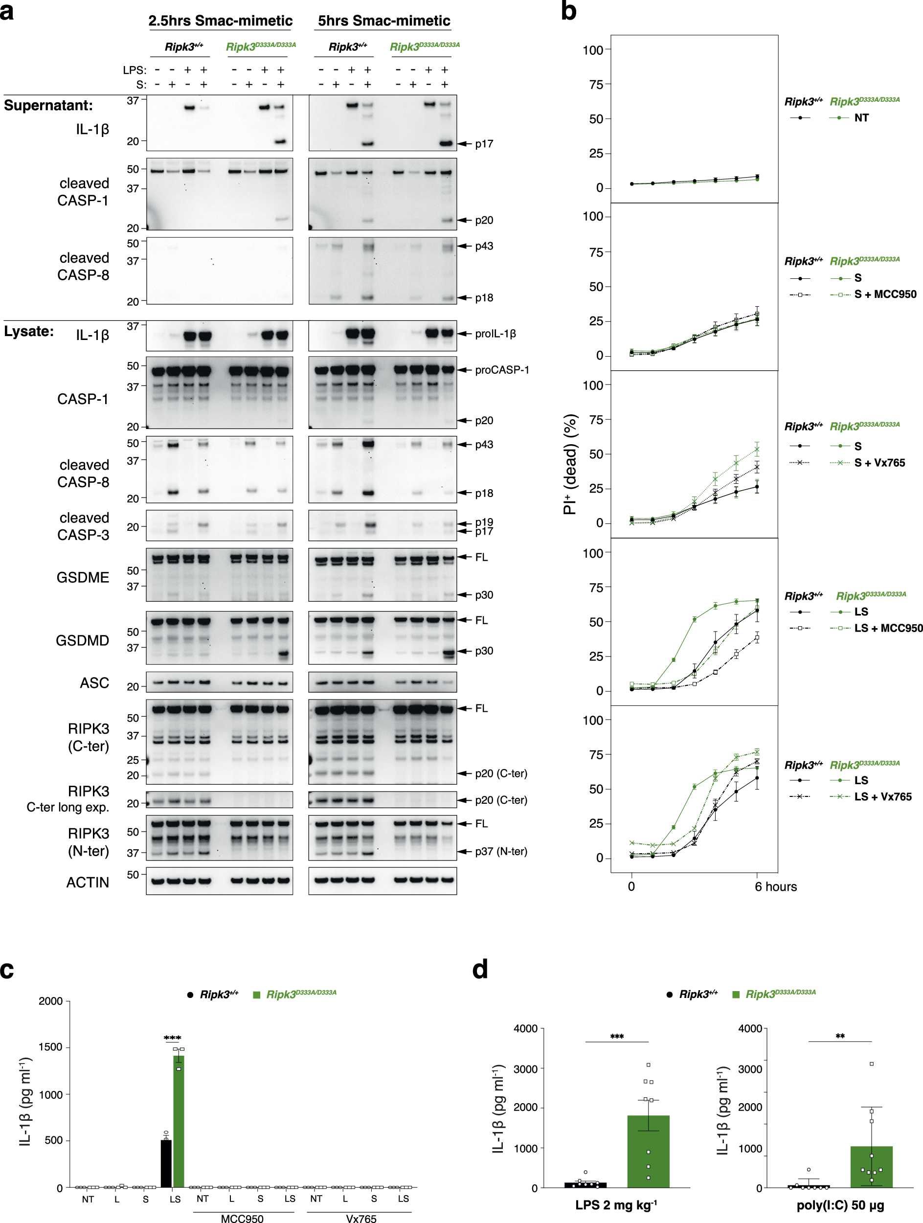
Conclusion: This cleavage does not prevent death; it restricts inflammation through negative feedback. This explains its high conservation—it regulates the intensity of inflammation, rather than whether a cell lives or dies.
III. Implications for experiments
- Establish "can be cleaved" and "cleavage is required" separately: First confirm that the bands are genuine cleavage fragments using three controls—wild type, mutant and inhibitor—so that a subsequent negative phenotype is meaningful.
- Actively rule out compensation when drawing a negative conclusion: Could normal RIPK1 cleavage mask the effect of the RIPK3 mutation? The double-mutant mice directly address this question.
- Use pathway-specific controls to define the scope of an effect: The response to compound A (acting through RIPK3) is amplified, while those to nigericin/TAK1i (not acting through RIPK3) remain unchanged; this set of controls can locate the step affected. Checking priming before discussing activation is fundamental to inflammasome research.
IV. Links to other papers on this site
- Extension | RIPK3 activates NLRP3 without MLKL (Lawlor 2015, Nat Commun): The Lawlor paper establishes the existence of the proinflammatory TLR–RIPK3–caspase-8–NLRP3 axis; this paper further shows that caspase-8 cleavage of RIPK3 is a negative feedback mechanism that restricts this axis.
- Extension | Mitochondrial apoptosis and NLRP3 (Allam 2014, EMBO Rep): From the same research group. The Allam paper finds that caspase-8 promotes inflammasome priming; this paper finds that it cleaves RIPK3 to restrict NLRP3 activation—caspase-8 regulates inflammation in both directions.
- Comparison | Caspase-1 self-cleavage terminates inflammasome activity (Boucher 2018, JEM): In the Boucher paper, caspase-1 self-cleavage limits the duration of inflammasome activity; in this paper, caspase-8 cleavage of RIPK3 limits the intensity of activation. Each of these two proinflammatory pathways has its own negative feedback mechanism.
- Comparison | TNF-α + IFN-γ synergistically induce PANoptosis (Karki 2021, Cell): In the Karki paper, caspase-8 drives cell death; in this paper, it restricts pyroptosis by cleaving RIPK3—with the same caspase-8, substrate choice determines the direction of the effect.
(The comparisons across papers above are integrative interpretations, rather than direct conclusions of any one original paper.)
What remains unresolved
Limitations (two): ① The actual substrate through which caspase-8 inhibits necroptosis has yet to be identified; ② the molecular mechanism of the negative feedback remains unclear, the in vivo readout is limited to serum IL-1β at 2 hours, and long-term inflammatory outcomes are unknown.
RIPK3 cleavage is dispensable for necroptosis inhibition but restricts NLRP3 inflammasome activation
Hong Tri Tran; Tobias Kratina; Auriane Coutansais; Dominika Michalek; Benjamin M. Hogan; Kate E. Lawlor; James E. Vince; John Silke; Najoua Lalaoui Cell Death & Differentiation. 2024;31:662-671.
Figures are from the verified original paper stored in the library. Original labels, axes and controls are retained; figure numbers and sources are registered in the library.
References
- Tran HT, Kratina T, Coutansais A, et al. RIPK3 cleavage is dispensable for necroptosis inhibition but restricts NLRP3 inflammasome activation. Cell Death Differ. 2024;31(5):662-671. doi:10.1038/s41418-024-01281-x
- Newton K, Wickliffe KE, Dugger DL, et al. Cleavage of RIPK1 by caspase-8 is crucial for limiting apoptosis and necroptosis. Nature. 2019;574(7778):428-431.
- Lalaoui N, Boyden SE, Oda H, et al. Mutations that prevent caspase cleavage of RIPK1 cause autoinflammatory disease. Nature. 2020;577(7788):103-108.
- Kang TB, Yang SH, Toth B, Kovalenko A, Wallach D. Caspase-8 blocks kinase RIPK3-mediated activation of the NLRP3 inflammasome. Immunity. 2013;38(1):27-40.
Paper discussion
No sign-up. Nickname optional.