- 01
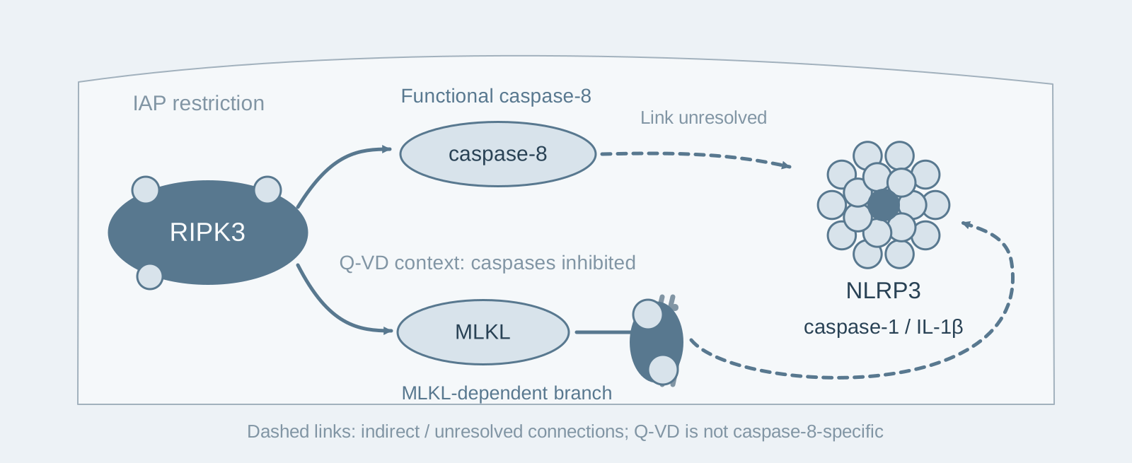
Main finding: When IAPs are inhibited, RIPK3 deficiency markedly reduces caspase-1 cleavage and IL-1β secretion, whereas MLKL deficiency has no effect;
- 02
Key mechanism: RIPK3 activates NLRP3 through caspase-8; the RIPK3–MLKL pathway takes over only after caspase-8 is inhibited;
- 03
Significance: RIPK3's pro-inflammatory function can be separated from necroptosis, so MLKL deletion cannot determine whether inflammation is driven by RIPK3.
On this page
1. What question does this paper answer?
Necroptosis is executed by RIPK3–MLKL, but RIPK3 has also been reported to promote NLRP3 inflammasome activation. This paper asks: Must RIPK3's pro-inflammatory activity act through MLKL, that is, through necroptosis? The answer is no: when IAPs are inhibited, RIPK3 mainly activates NLRP3 through caspase-8, and this pathway does not require MLKL.
2. Following the chain of evidence
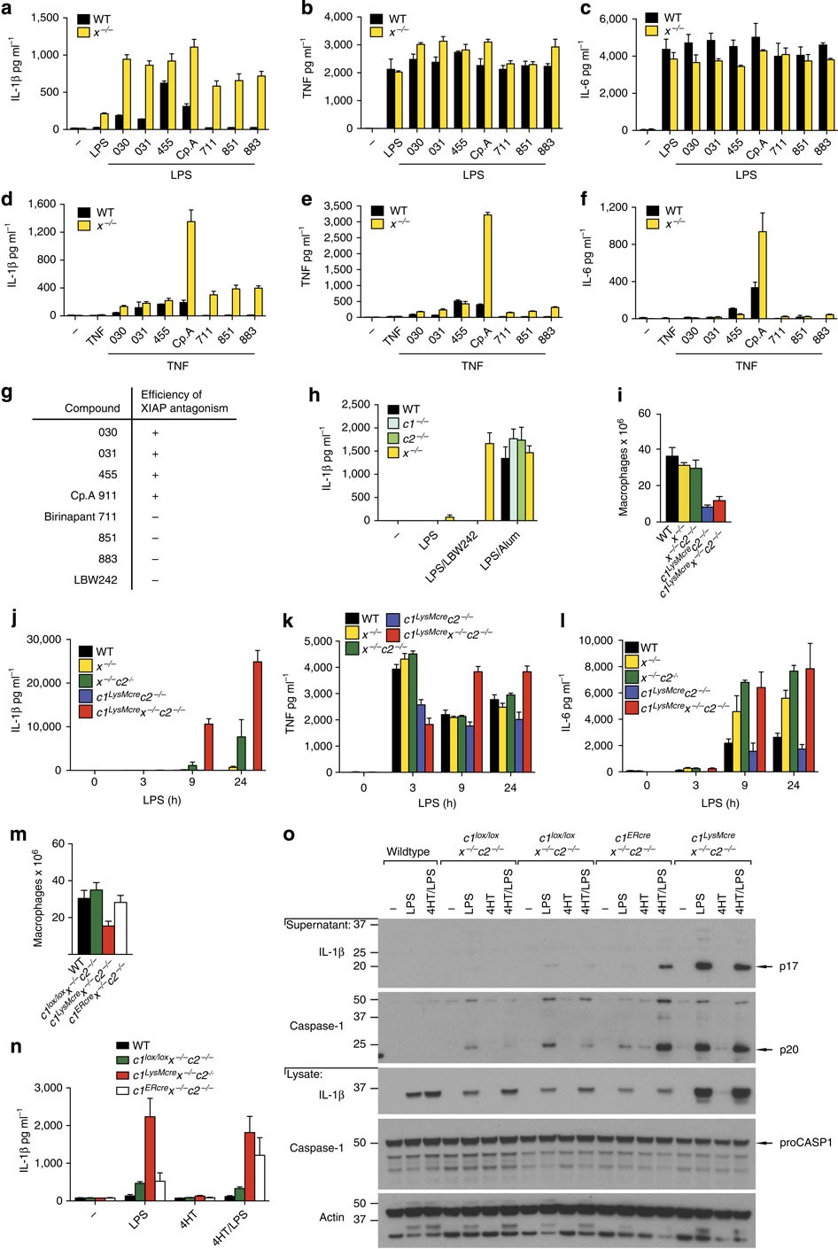
Step 1: Identify which IAP limits IL-1β secretion (Fig. 1). The authors treated LPS- or TNF-primed mouse bone marrow-derived macrophages (BMDMs) with Smac-mimetic compounds of different selectivities. Only bivalent compounds that also antagonized XIAP, such as Compound A, induced IL-1β secretion; compounds targeting only cIAP1/2 did not. Genetic deletion gave consistent results: deleting XIAP alone had some effect, while macrophages lacking all three proteins—XIAP, cIAP1 and cIAP2—secreted the most IL-1β and IL-18. These results show that XIAP is a key IAP limiting IL-1β secretion and that the three IAPs have functionally redundant roles.
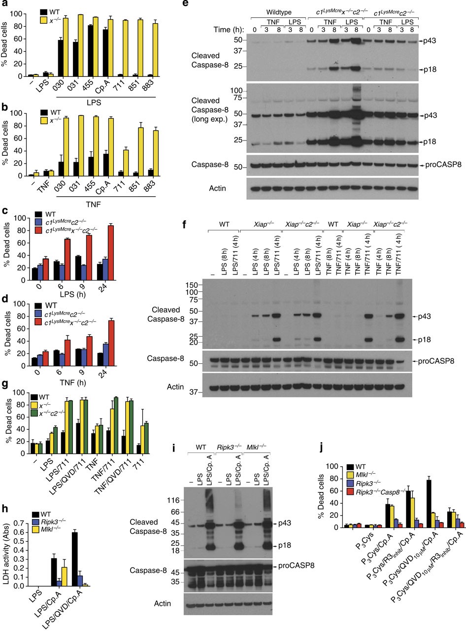
Step 2: Distinguish the roles of RIPK3 and MLKL in cell death (Fig. 2). In XIAP-deficient BMDMs, the authors additionally deleted either RIPK3 or MLKL and stimulated the cells with LPS or TNF. RIPK3 deficiency reduced caspase-8 modification and apoptosis while also blocking necroptosis; MLKL deficiency blocked only necroptosis and did not affect apoptosis. These results show that RIPK3 lies upstream of the branch point and promotes both apoptosis and necroptosis, whereas MLKL is responsible only for necroptosis.
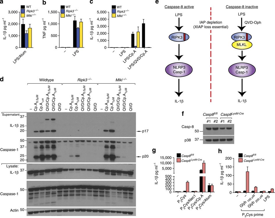
Step 3: Test whether RIPK3 requires MLKL to activate NLRP3 (Figs. 3–5). The authors treated wild-type, Ripk3−/− and Mlkl−/− BMDMs with LPS plus Compound A. Caspase-1 cleavage and IL-1β secretion were markedly impaired in Ripk3−/− cells, whereas Mlkl−/− cells were indistinguishable from wild-type cells. After addition of the caspase-8 inhibitor Q-VD, however, inflammasome activation became dependent on MLKL. These results show that RIPK3 activates NLRP3 through caspase-8 without requiring MLKL; the RIPK3–MLKL pathway takes over only when caspase-8 function is inhibited. Further treatment with the RIPK3 kinase inhibitor GSK872 showed that, as long as caspase-8 function remained intact, inhibiting RIPK3 kinase activity did not affect IL-1β secretion, indicating that RIPK3 mainly acts as a scaffold in this setting. The authors also generated Ripk3−/−Casp8−/− double-knockout cells, which did not respond at all to Compound A-induced inflammasome activation but responded normally to nigericin. This shows that the pathway specifically depends on RIPK3 and caspase-8, while the canonical NLRP3 activation mechanism itself remains intact.
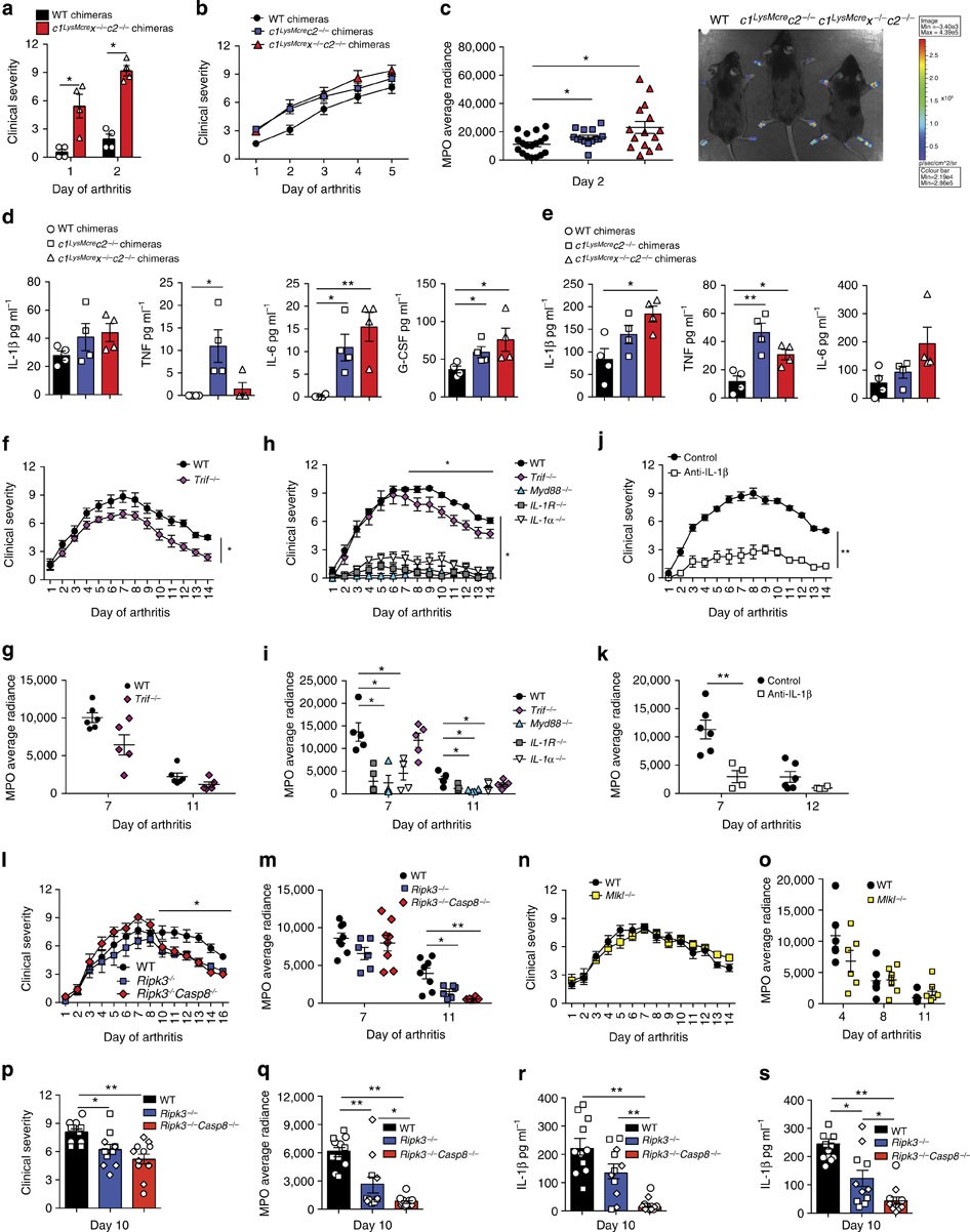
Step 4: Validate the findings in vivo (Figs. 8–9). The authors observed spontaneous arthritis in mice lacking IAPs in myeloid cells, with elevated serum IL-1β, IL-6 and TNF; treatment with a TNF-neutralizing antibody alleviated disease. In the K/B×N serum-transfer arthritis model, joint inflammation resolved more rapidly and local joint IL-1β was lower in Ripk3−/− mice, whereas disease progression in Mlkl−/− mice did not differ from wild-type mice. These results show that RIPK3's MLKL-independent pro-inflammatory function also exists in inflammatory disease in vivo.
3. Implications for experiments
- MLKL deletion cannot determine "whether inflammation is driven by RIPK3": In this study, the phenotype was normal in Mlkl−/− cells but markedly impaired in Ripk3−/− cells. The separation of these phenotypes itself provides evidence that RIPK3 uses an MLKL-independent pathway.
- When choosing a Smac mimetic, check whether it antagonizes XIAP: Compounds targeting only cIAP1/2 did not induce IL-1β secretion, so choosing the wrong compound would produce a negative result.
- Assess how RIPK3 acts in conjunction with caspase-8 inhibition: Adding Q-VD switched the pathway from MLKL-independent to MLKL-dependent; without the inhibitor, this relationship would be missed.
4. Connections with other papers on this site
- Comparison | BAX/BAK-mediated mitochondrial apoptosis activates IL-1β (Vince 2018, Cell Rep): In the same IAP-deficient model, Vince's paper emphasizes the BAX/BAK–caspase-3/7 apoptotic axis driving IL-1β, whereas this paper emphasizes the RIPK3–caspase-8–NLRP3 axis. The two pathways complement each other in the same setting.
- Extension | RIPK3 cleavage limits NLRP3 (Tran 2024, Cell Death Differ): This paper establishes the existence of the pro-inflammatory TLR–RIPK3–caspase-8–NLRP3 axis. Tran's paper further shows that caspase-8 cleavage of RIPK3 provides negative feedback limiting that axis.
- Extension | Mitochondrial apoptosis and NLRP3 (Allam 2014, EMBO Rep): Allam's paper reports that caspase-8 participates in inflammasome priming, while this paper reports its direct involvement in NLRP3 activation. Together, they show that caspase-8 has multiple pro-inflammatory functions beyond apoptosis.
- Extension | TLR-induced death of IFN-γ-primed macrophages (Simpson 2022, Immunity): The same research group conducted both studies. In this paper, caspase-8 activates NLRP3 through RIPK3; in Simpson's paper, caspase-8 lowers BCL-2 through transcriptional regulation to drive mitochondrial apoptosis—further expanding its noncanonical functions.
(The cross-paper comparisons above are a synthesis, rather than direct conclusions of any one original paper.)
What remains unresolved
Limitations: ① The specific molecular pathway through which RIPK3 increases IL-1β in the in vivo arthritis models was not directly established; ② the mechanistic experiments were mainly performed in mouse macrophages, and the corresponding relationships in human cells were not tested.
RIPK3 promotes cell death and NLRP3 inflammasome activation in the absence of MLKL
Kate E Lawlor; Nufail Khan; Alison Mildenhall; Motti Gerlic; Ben A Croker; Akshay A D’Cruz; Cathrine Hall; Sukhdeep Kaur Spall; Holly Anderton; Seth L Masters; Maryam Rashidi; Ian P Wicks; Warren S Alexander; Yasuhiro Mitsuuchi; Christopher A Benetatos; Stephen M Condon; W Wei-Lynn Wong; John Silke; David L Vaux; James E Vince Nature Communications. 2015;6:.
Figures are from the verified original paper stored in the library. Original labels, axes and controls are retained; figure numbers and sources are registered in the library.
References
- Lawlor KE, Khan N, Mildenhall A, et al. RIPK3 promotes cell death and NLRP3 inflammasome activation in the absence of MLKL. Nat Commun. 2015;6:6282. doi:10.1038/ncomms7282
- Vince JE, De Nardo D, Gao W, et al. The mitochondrial apoptotic effectors BAX/BAK activate caspase-3 and -7 to trigger NLRP3 inflammasome and caspase-8 driven IL-1β activation. Cell Rep. 2018;25(9):2339-2353.e4.
- Murphy JM, Czabotar PE, Hildebrand JM, et al. The pseudokinase MLKL mediates necroptosis via a molecular switch mechanism. Immunity. 2013;39(3):443-453.
Paper discussion
No sign-up. Nickname optional.