- 01
Main finding: BAX/BAK-mediated mitochondrial apoptosis is not "silent": it activates caspase-1 and IL-1β, while TNF secretion remains unaffected;
- 02
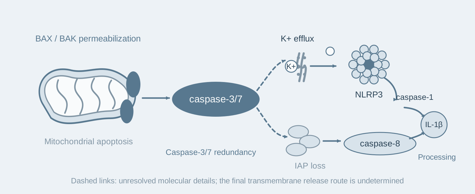
Key mechanism: Caspase-3/7 simultaneously drives two pathways: one activates NLRP3 through potassium efflux, while the other activates caspase-8 through IAP loss to directly cleave IL-1β;
- 03
Significance: The study revises the idea that "apoptosis is necessarily immunologically silent" and provides a mechanistic explanation for inflammation accompanying pro-apoptotic anticancer drugs.
On this page
1. What question does this paper answer?
The traditional view considers mitochondrial apoptosis immunologically silent: apoptotic cells do not immediately rupture their membranes and are cleared by phagocytes. However, death receptor- and TLR-associated apoptosis is not always quiet: caspase-8 can sometimes directly cleave IL-1β. Vince and colleagues therefore asked a specific question: Can BAX/BAK-mediated mitochondrial apoptosis activate IL-1β in macrophages? If so, is this carried out by NLRP3, caspase-8, or cell lysis caused by gasdermin pore formation?
2. Following the logic of the evidence
Step 1: Establishing a system that specifically initiates BAX/BAK apoptosis (Figure 1). The authors inhibited two anti-apoptotic proteins separately and together in macrophages. Inhibiting BCL-XL alone did not kill the cells; rapid death occurred only when MCL-1 was also depleted. Cells with a Bax/Bak double knockout resisted all the combinations. These results show that death induced by this system specifically proceeds through BAX/BAK, and all subsequent conclusions are based on this system.
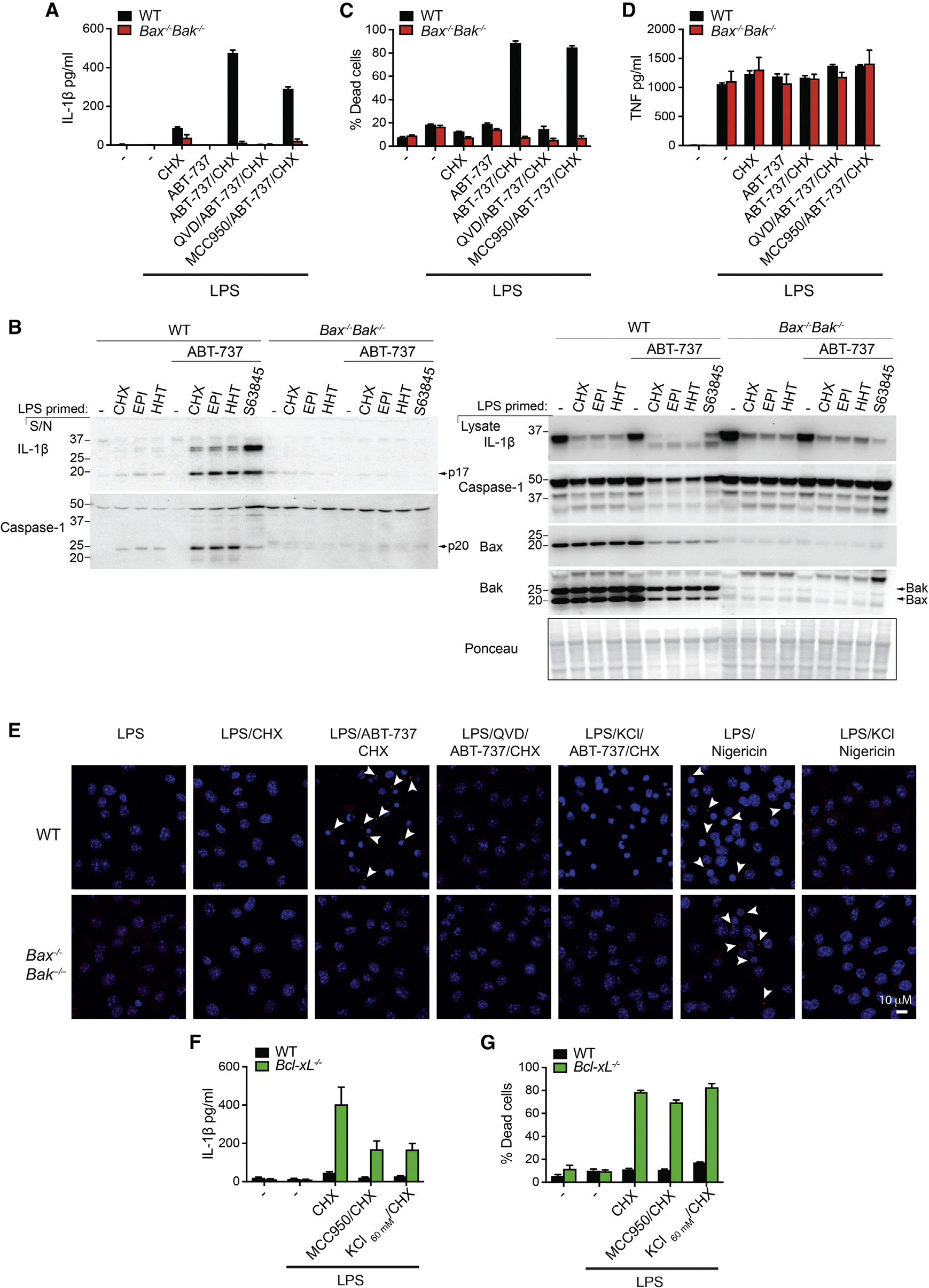
Step 2: Confirming the inflammatory response and positioning upstream and downstream events (Figures 2–3). The authors first primed cells with LPS and then initiated BAX/BAK apoptosis. Caspase-1 and IL-1β were markedly activated, while TNF secretion remained unchanged: the cells' ability to produce and secrete proteins was not impaired, and the affected pathway was specifically the IL-1β pathway. The authors then treated cells with high potassium and MCC950. Both inhibited IL-1β but could not prevent apoptosis. These results show that cell death and inflammatory activation are two separable events, with NLRP3 downstream of BAX/BAK and dependent on potassium efflux.
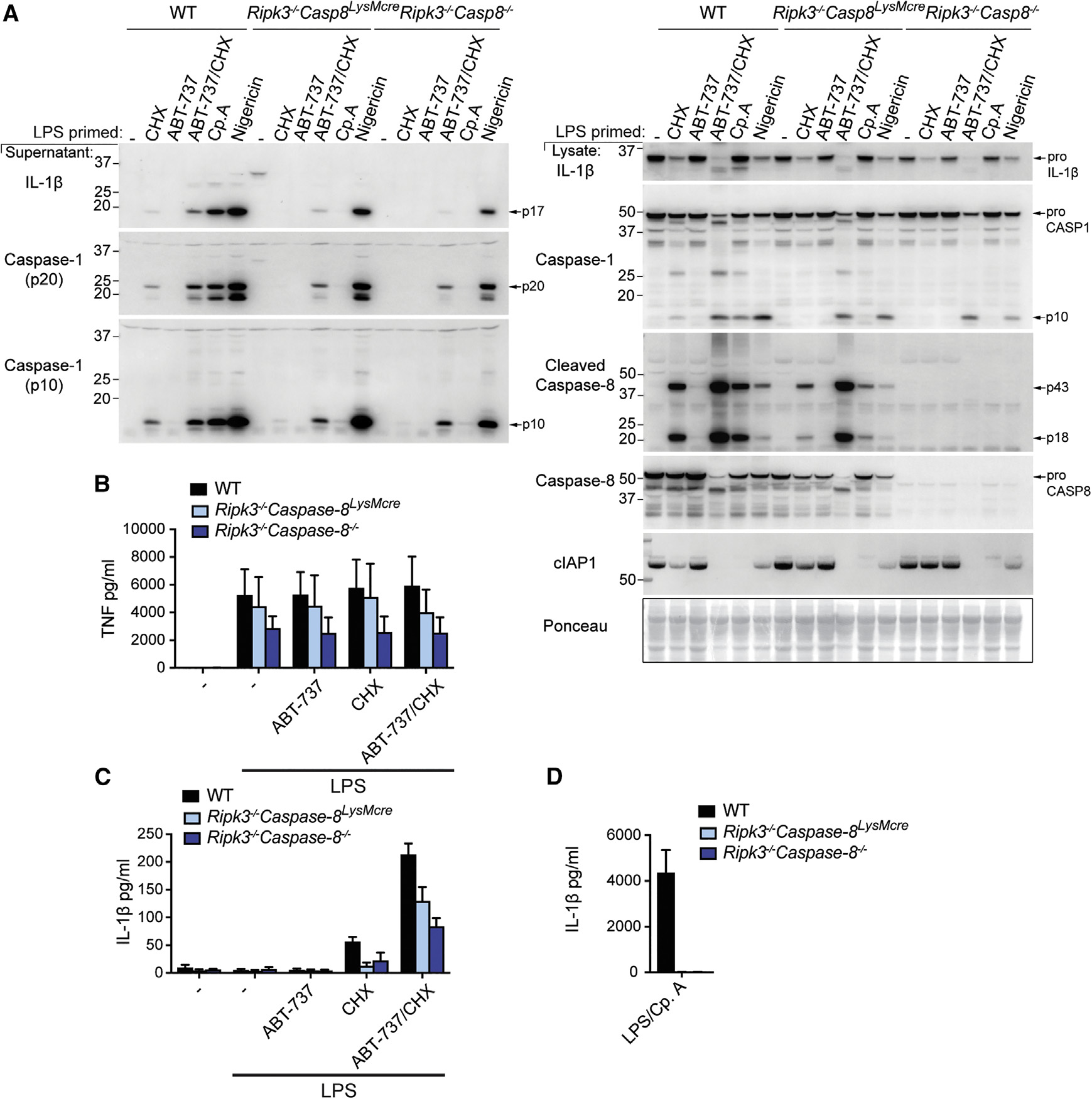
Step 3: Distinguishing two parallel pathways (Figures 4–6). The authors knocked out NLRP3 or caspase-1 and found that IL-1β was only partially reduced: the canonical pathway could not account for all of it. Although GSDMD was cleaved, knocking it out did not affect IL-1β release. The authors then examined caspase-8. BAX/BAK activation was accompanied by IAP protein loss and caspase-8 activation, which also occurred without LPS, excluding interference from TLR stimulation. IL-1β was markedly reduced in caspase-8-deficient cells, while caspase-1 activation continued as usual. These results show that caspase-8 forms a second IL-1β-processing pathway parallel to NLRP3, rather than acting upstream of NLRP3.
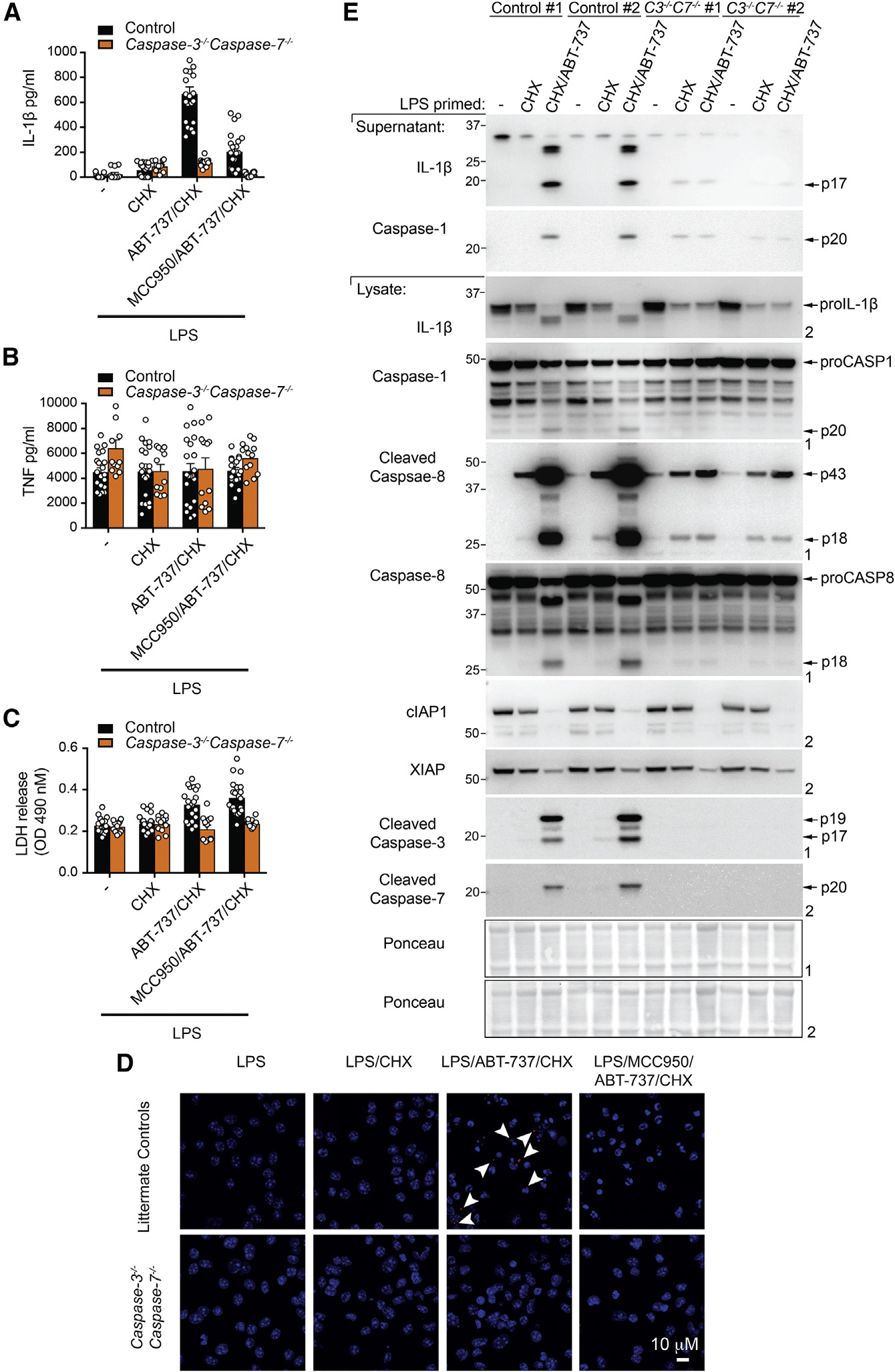
Step 4: Identifying the common upstream component (Figure 7). Knocking out caspase-3 alone produced no phenotype. Only the caspase-3/7 double knockout blocked both pathways simultaneously, while TNF secretion and the response to nigericin remained normal. GSDME was likewise cleaved and likewise dispensable. These results show that caspase-3/7 is functionally redundant and is the common upstream component linking apoptosis to inflammation.
Conclusion: After BAX/BAK activation, caspase-3/7 activates NLRP3 through potassium efflux along one pathway and activates caspase-8 through IAP loss to directly cleave IL-1β along the other; gasdermin cleavage is merely an accompanying event.
3. Implications for experiments
- Assess death and inflammation separately: Inhibiting the inflammasome does not mean saving the cells. The measurement panel should include death readouts (PI/LDH), cleavage of the different caspases, ASC specks, extracellular and intracellular IL-1β, and a control involving an unrelated cytokine such as TNF.
- "Cleaved" does not mean "required": Both GSDMD and GSDME are cleaved, yet neither knockout affects the outcome. Cleavage is only a clue to involvement; knockout is needed to determine the functional role.
- Redundancy requires a double knockout to reveal it: A caspase-3 single knockout has no phenotype, which can easily lead to the mistaken conclusion that it is "unimportant". Double knockouts should be considered for executor molecules in the same family.
4. Connections to other papers on this site
- Comparison | Mitochondrial apoptosis and NLRP3 (Allam 2014, EMBO Rep): The Allam paper shows that canonical NLRP3 stimuli do not require mitochondrial apoptosis. This paper shows that an apoptotic system in which BCL-XL and MCL-1 are both inhibited can indeed drive IL-1β. Whether something is "required" must be stated together with the stimulus context; the two papers do not contradict each other.
- Comparison | RIPK3 activates NLRP3 without requiring MLKL (Lawlor 2015, Nat Commun): Within the same context of limited IAP activity, the Lawlor paper emphasizes the RIPK3–caspase-8–NLRP3 axis, while this paper emphasizes the BAX/BAK–caspase-3/7 axis. The two pathways complement each other.
- Comparison | GSDMD is an executor of pyroptosis (He 2015, Cell Res): The He paper establishes the central role of the caspase-1–GSDMD axis in canonical inflammasome signaling. This paper shows that GSDMD is cleaved during BAX/BAK apoptosis but is not required: the cell-death context determines the release mechanism.
- Extension | TLR-induced death in IFN-γ-primed macrophages (Simpson 2022, Immunity): This paper shows that MCL-1 and BCL-XL jointly restrain BAX/BAK in macrophages. The Simpson paper shows that IFN-γ/LPS reduces BCL-2 through caspase-8 and reduces MCL-1 through iNOS, releasing this restraint from two directions.
(The comparisons across papers above are a synthesis, rather than a direct conclusion of any one original paper.)
What remains unresolved
Limitations (two points): ① How caspase-3/7 causes potassium efflux, and the route by which IL-1β ultimately crosses the membrane for release, were not determined. ② How BAX/BAK causes IAP degradation remains unclear, and the evidence comes primarily from an in vitro mouse macrophage system.
The Mitochondrial Apoptotic Effectors BAX/BAK Activate Caspase-3 and -7 to Trigger NLRP3 Inflammasome and Caspase-8 Driven IL-1β Activation
James E. Vince; Dominic De Nardo; Wenqing Gao; Angelina J. Vince; Cathrine Hall; Kate McArthur; Daniel Simpson; Swarna Vijayaraj; Lisa M. Lindqvist; Philippe Bouillet; Mark A. Rizzacasa; Si Ming Man; John Silke; Seth L. Masters; Guillaume Lessene; David C. S. Huang; Daniel H. D. Gray; Benjamin T. Kile; Feng Shao; Kate E. Lawlor Cell Reports. 2018;25:2339-2353.e4.
Figures are from the verified original paper stored in the library. Original labels, axes and controls are retained; figure numbers and sources are registered in the library.
References
- Vince JE, De Nardo D, Gao W, et al. The Mitochondrial Apoptotic Effectors BAX/BAK Activate Caspase-3 and -7 to Trigger NLRP3 Inflammasome and Caspase-8 Driven IL-1β Activation. Cell Reports. 2018;25(9):2339-2353.e4. doi:10.1016/j.celrep.2018.10.103
- He WT, Wan H, Hu L, et al. Gasdermin D is an executor of pyroptosis and required for interleukin-1β secretion. Cell Research. 2015;25(12):1285-1298.
- Lawlor KE, Feltham R, Yabal M, et al. XIAP loss triggers RIPK3- and caspase-8-driven IL-1β activation and cell death as a consequence of TLR-MyD88-induced cIAP1-TRAF2 degradation. Cell Reports. 2017;20(3):668-682.
Paper discussion
No sign-up. Nickname optional.