- 01
Main finding: IFN-γ priming sensitizes macrophages to TLR ligand-induced death, which depends on caspase-8 and BAX/BAK;
- 02
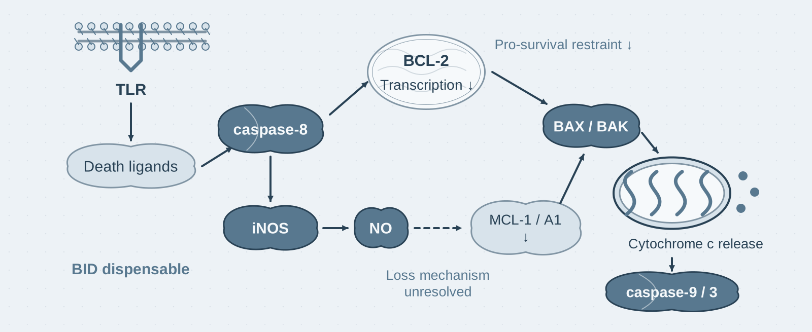
Key mechanism: Caspase-8 lowers BCL-2 through transcriptional regulation, and iNOS/NO lowers MCL-1; together, these changes remove the restraints on BAX/BAK;
- 03
Significance: HLH lethality depends on caspase-8 rather than iNOS; during SARS-CoV-2 infection, iNOS and caspase-8 worsen disease without changing viral load.
On this page
I. What question does this paper answer?
During infection, macrophages encounter both IFN-γ and pathogen-derived TLR ligands. What happens to cell fate when these two signals are combined? This paper asks: Does IFN-γ priming change the macrophage response to TLR stimulation, and if it causes death, which pathway does it use? The conclusion is that IFN-γ priming sensitizes macrophages to TLR ligand-induced death. Death ligands (TNF and FASL) initiate caspase-8 signaling, which, through transcriptional regulation and iNOS/NO, removes the restraints imposed by the BCL-2 family on BAX/BAK and ultimately kills the cells through mitochondrial apoptosis.
II. Tracing the argument
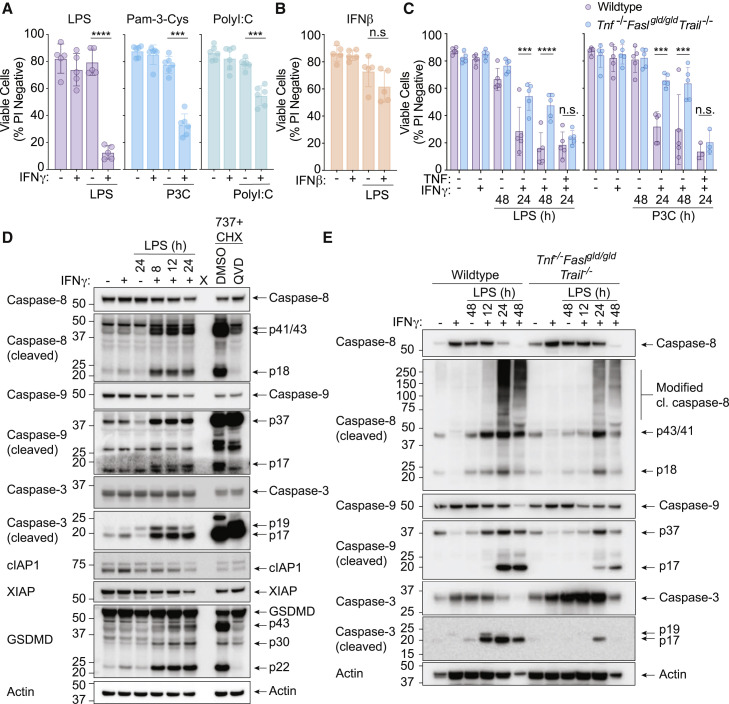
Step 1: Confirm the phenomenon and identify the triggers of death (Figure 1). The authors primed mouse bone marrow-derived macrophages (BMDMs) with IFN-γ overnight and then added LPS, Pam3CSK4, or poly(I:C). All three TLR ligands killed the cells within 24 hours, whereas IFN-β priming had no such effect; IFN-γ alone or LPS alone was also nonlethal. To identify the triggers of death, the authors examined the contribution of death ligands and found that cells lacking all three signaling systems—TNF, FASL, and TRAIL—were markedly resistant to IFN-γ/LPS killing, while adding exogenous TNF restored death. Caspase-8, -9, and -3 were cleaved and activated in these cells, and GSDMD was also cleaved, but deletion of GSDMD did not affect death. These results show that TLR-induced death after IFN-γ priming is initiated by death ligands and is not executed by GSDMD-mediated pyroptosis.
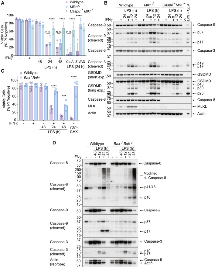
Step 2: Locate the execution pathway using gene deletion (Figures 2–3). The authors repeated the treatment in BMDMs lacking both Casp8 and Mlkl and found that these cells were largely resistant to IFN-γ/LPS killing. Cells lacking both Bax and Bak were also substantially resistant, although less so than cells lacking caspase-8. The authors then measured mitochondrial cytochrome c retention and found that WT cells lost cytochrome c over time, whereas caspase-8-deficient cells did not. Deletion of BID, a canonical caspase-8 substrate, had no effect on cell death, cytochrome c release, or caspase-3/9 activation. These results show that IFN-γ/LPS killing is mediated by caspase-8-driven BAX/BAK mitochondrial apoptosis and does not proceed through BID.
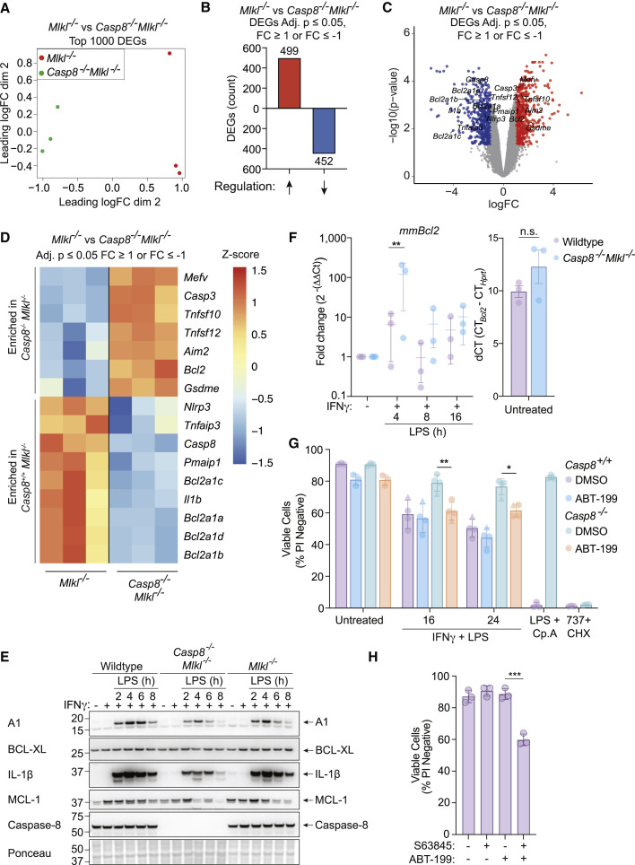
Step 3: Determine how caspase-8 connects to mitochondria (Figures 4–5). The authors performed transcriptome sequencing on caspase-8-deficient and control cells and found that loss of caspase-8 altered the expression of many genes; among these changes, increased pro-apoptotic NOXA and decreased pro-survival Bcl2 were associated with BAX/BAK activation. Treating caspase-8-deficient cells with the clinically approved BCL-2 inhibitor venetoclax partially restored IFN-γ/LPS killing; simultaneous inhibition of BCL-2 and MCL-1 killed macrophages without any infection-associated signal. The authors further tested knock-in mice carrying catalytically inactive caspase-8 (C362S) and found that loss of enzymatic activity provided protection approaching that of complete deletion. These results show that caspase-8 uses its catalytic activity to suppress BCL-2 at the transcriptional level, which, together with loss of MCL-1, removes the restraints on BAX/BAK.
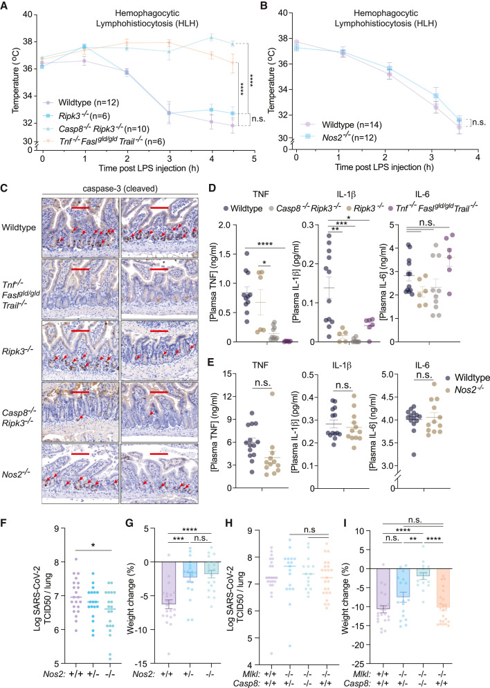
Step 4: Establish iNOS/NO as a prerequisite and test the pathway in vivo (Figures 6–7). The authors treated cells with the iNOS inhibitor 1400W or deleted Nos2 and found that both interventions blocked IFN-γ/LPS-induced death and cytochrome c release; adding the nitric oxide donor SNAP restored sensitivity. The role of iNOS was to reduce MCL-1 and A1 protein levels. In vivo, in a poly(I:C)+LPS-induced HLH (hemophagocytic lymphohistiocytosis) model, mice lacking caspase-8 or all three death-ligand signaling systems were protected, whereas Nos2-deficient mice developed disease just as WT mice did. In a nonlethal SARS-CoV-2 infection model, deletion of either Nos2 or caspase-8 reduced weight loss, but neither changed viral load in the lungs. These results show that the pathway affects disease severity in vivo rather than antiviral capacity, and that HLH lethality does not depend on iNOS.
III. Implications for experiments
- "Priming order" is a variable in experimental design: LPS given before IFN-γ, or both added simultaneously, did not cause death; only overnight IFN-γ priming followed by a TLR ligand was lethal—the order of stimulation and the interval between stimuli must be recorded in the methods and kept consistent.
- Compare the degree of resistance in knockout phenotypes: Resistance in Bax/Bak-double-deficient cells was weaker than in caspase-8-deficient cells, suggesting that caspase-8 has additional pro-death functions outside BAX/BAK—the relationship between the two pathways is "partial overlap" rather than "upstream and downstream".
- A broad-spectrum caspase inhibitor is not equivalent to caspase deletion: In this paper, IDN-6556 did not fully block IFN-γ/LPS killing, and BAX/BAK-mediated mitochondrial damage remained lethal when downstream caspases were inhibited—a negative inhibitor result does not rule out mitochondrial apoptosis.
IV. Connections to other papers on this site
- Comparison | TNF-α + IFN-γ synergistically induces PANoptosis (Karki 2021, Cell): Both papers examine macrophage death involving IFN-γ, iNOS, and caspase-8. Karki proposed iNOS/NO as the lethal mechanism and suggested its involvement in HLH; this paper tested that directly in gene-knockout mice and found that HLH lethality depends on caspase-8 but not iNOS—the in vivo conclusions need to be considered separately.
- Extension | BAX/BAK mitochondrial apoptosis activates IL-1β (Vince 2018, Cell Rep): Vince showed that MCL-1 and BCL-XL jointly restrain BAX/BAK in macrophages; this paper further shows that IFN-γ/LPS suppresses BCL-2 through caspase-8 and lowers MCL-1 through iNOS, removing these restraints from two directions.
- Extension | RIPK3 activates NLRP3 without MLKL (Lawlor 2015, Nat Commun) and Mitochondrial apoptosis and NLRP3 (Allam 2014, EMBO Rep): Caspase-8's non-apoptotic function is inflammasome priming in Allam and NLRP3 activation in Lawlor, whereas here it regulates transcription of BCL-2 family genes—further extending the range of caspase-8's pro-inflammatory functions.
(The comparisons across papers above are integrated interpretations, not direct conclusions of any single original paper.)
What remains unresolved
Limitations: (1) The molecular mechanism by which iNOS/NO lowers MCL-1 has not been defined; (2) the corresponding relationship in human macrophages was not tested, and the SARS-CoV-2 model was nonlethal, limiting its correspondence to severe COVID-19.
Interferon-γ primes macrophages for pathogen ligand-induced killing via a caspase-8 and mitochondrial cell death pathway
Daniel S. Simpson; Jiyi Pang; Ashley Weir; Isabella Y. Kong; Melanie Fritsch; Maryam Rashidi; James P. Cooney; Kathryn C. Davidson; Mary Speir; Tirta M. Djajawi; Sebastian Hughes; Liana Mackiewicz; Merle Dayton; Holly Anderton; Marcel Doerflinger; Yexuan Deng; Allan Shuai Huang; Stephanie A. Conos; Hazel Tye; Seong H. Chow; Arfatur Rahman; Raymond S. Norton; Thomas Naderer; Sandra E. Nicholson; Gaetan Burgio; Si Ming Man; Joanna R. Groom; Marco J. Herold; Edwin D. Hawkins; Kate E. Lawlor; Andreas Strasser; John Silke; Marc Pellegrini; Hamid Kashkar; Rebecca Feltham; James E. Vince Immunity. 2022;55:423–441.e9.
Figures are from the verified original paper stored in the library. Original labels, axes and controls are retained; figure numbers and sources are registered in the library.
References
- Simpson DS, Pang J, Weir A, et al. Interferon-γ primes macrophages for pathogen ligand-induced killing via a caspase-8 and mitochondrial cell death pathway. Immunity. 2022;55(3):423-441.e9. doi:10.1016/j.immuni.2022.01.003
- Karki R, Sharma BR, Tuladhar S, et al. Synergism of TNF-α and IFN-γ triggers inflammatory cell death, tissue damage, and mortality in SARS-CoV-2 infection and cytokine shock syndromes. Cell. 2021;184(1):149-168.e17.
- Vince JE, De Nardo D, Gao W, et al. The mitochondrial apoptotic effectors BAX/BAK activate caspase-3 and -7 to trigger NLRP3 inflammasome and caspase-8 driven IL-1β activation. Cell Rep. 2018;25(9):2339-2353.e4.
- Fritsch M, Günther SD, Schwarzer R, et al. Caspase-8 is the molecular switch for apoptosis, necroptosis and pyroptosis. Nature. 2019;575(7784):683-687.
Paper discussion
No sign-up. Nickname optional.