- 01
Main finding: Of the 8 candidate factors, only TNF-α and IFN-γ synergistically induce PANoptosis, and the resulting mouse shock model reproduces cytokine shock syndromes;
- 02
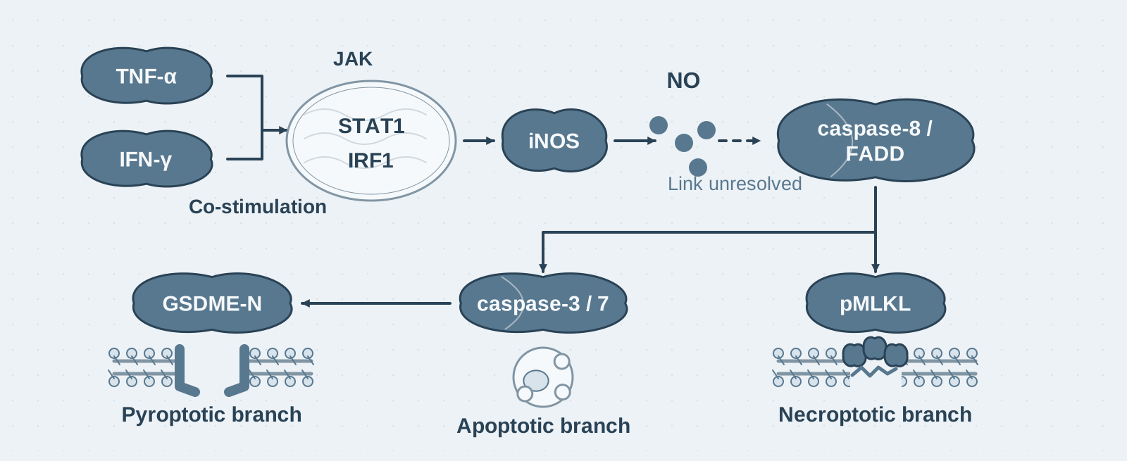
Key mechanism: The JAK–STAT1–IRF1 axis induces iNOS to produce sufficient NO, while caspase-8/FADD drives all three cell-death pathways simultaneously;
- 03
Significance: Neutralizing the two factors protects mice from four lethal models—giving cytokine storm a molecular definition that can be targeted for the first time.
On this page
I. What question does this paper answer?
Severe COVID-19 is associated with a "cytokine storm," but "storm" is only a clinical descriptive term: more than a dozen factors rise simultaneously in patient serum, and it remains unclear which combination, acting through which mechanism, turns inflammation into tissue damage and death. This paper asks: Is there a specific combination of factors within the storm that drives inflammatory cell death (PANoptosis: simultaneous activation of pyroptosis, apoptosis, and necroptosis), and can it become a therapeutic target?
II. Tracing the argument
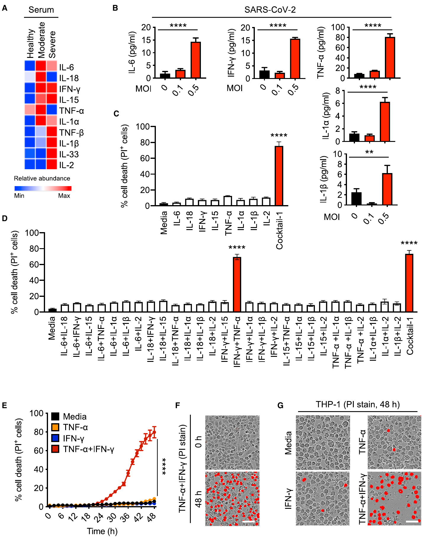
Step 1: Find the lethal combination of factors (Figure 1). Using public patient datasets, the authors selected 8 candidate factors and treated macrophages with each separately. None caused cell death, whereas a mixture of all 8 factors caused substantial death. The authors tested all 28 pairwise combinations, and only TNF-α + IFN-γ reached the level of cell death induced by the full mixture; even increasing the concentration of a mixture lacking these two factors by 10-fold did not cause death, indicating that the lethal effect specifically arose from their synergy. Patient data also showed that both factors were simultaneously elevated during severe disease.
Step 2: Show that this combination is sufficient to cause disease (Figure 2). Killing cells in vitro does not establish that a stimulus causes disease in vivo. The authors injected mice with TNF-α or IFN-γ separately and found that neither alone was lethal, whereas combined injection was synergistically lethal; 5 hours after injection, mice displayed findings consistent with severe COVID-19: intestinal and lung tissue damage, elevated serum LDH/ALT/ferritin, and reduced platelet counts. These results show that the two cytokines themselves are sufficient to cause tissue damage and death, rather than merely accompanying disease.
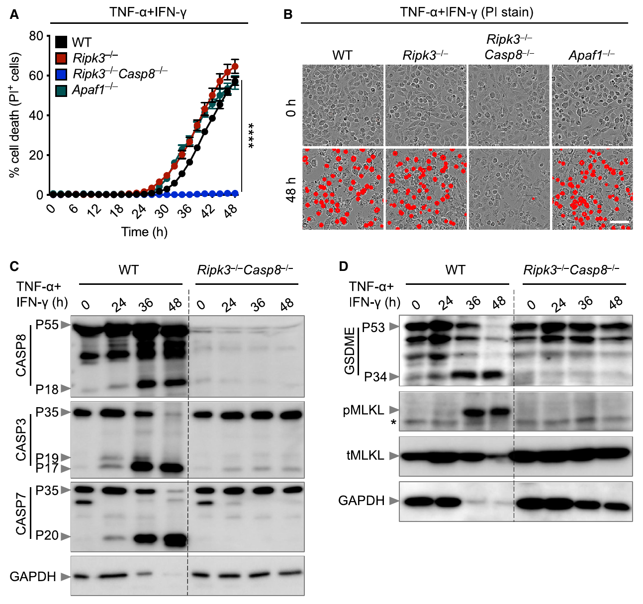
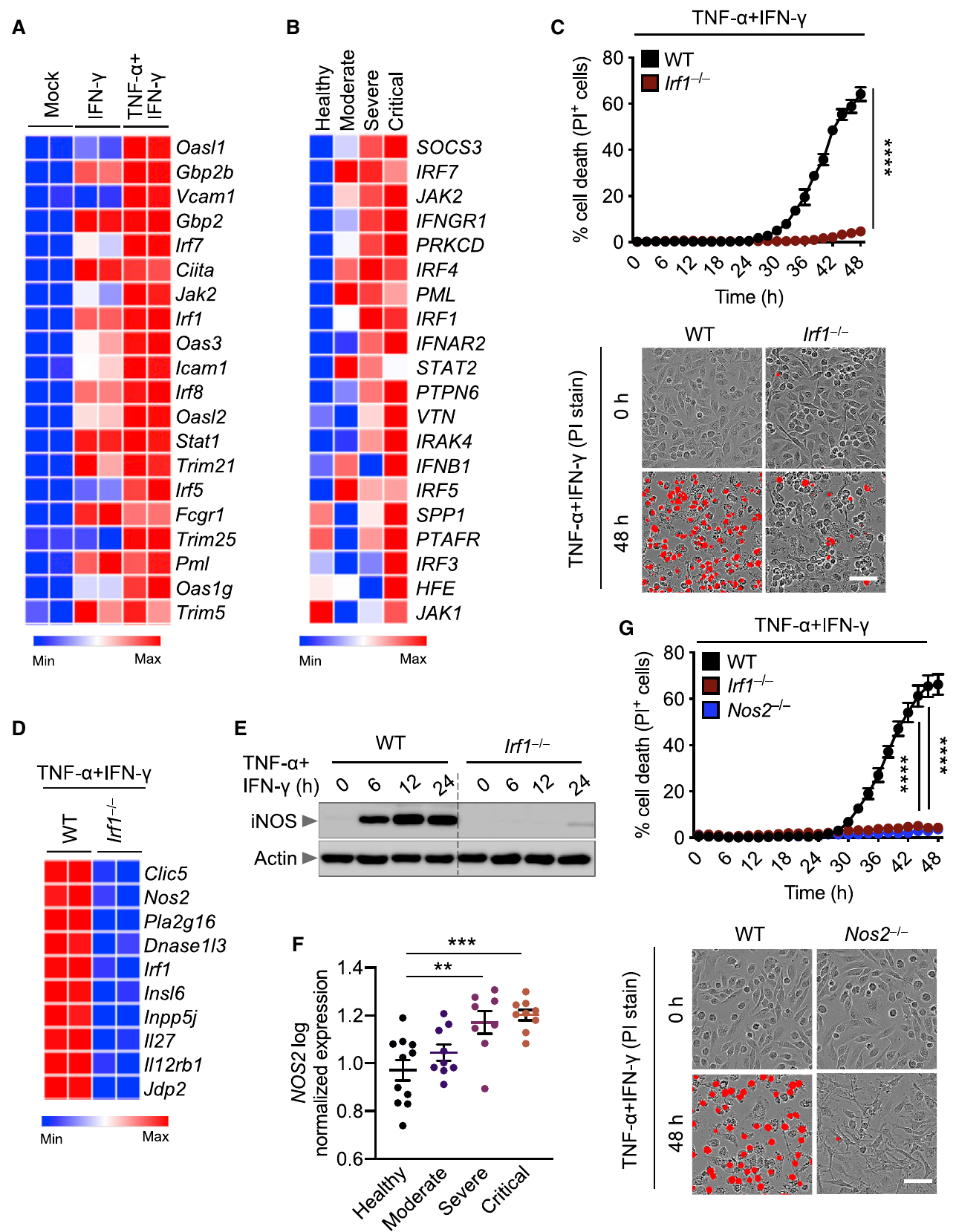
Step 3: Identify the type and mechanism of cell death (Figures 3–5). In cells receiving the combined treatment, the authors detected molecular markers of three cell-death pathways simultaneously: GSDME cleavage, caspase-3/7/8 cleavage, and MLKL phosphorylation; caspase-1 was not activated, while GSDMD was instead cleaved into an inactive P20 fragment—this death was PANoptosis rather than canonical pyroptosis. Upstream, the authors screened cells lacking 17 different interferon-pathway genes and found that only deletion of STAT1 or IRF1 protected cells; deletion of the downstream iNOS or treatment with NO inhibitors also blocked death. These results show that the basis of the synergy is an NO concentration threshold: the amount of NO induced by either factor alone is insufficient to cause death, and only their combination exceeds the threshold. At the downstream execution stage, individual deletion of GSDME, MLKL, or caspase-3 only partially reduced death, whereas only deletion of caspase-8 or FADD provided complete protection. The three downstream pathways are mutually redundant, and caspase-8/FADD is their common upstream component.
Step 4: In vivo validation and treatment (Figures 6–7). The authors tested knockout mice in the cytokine-shock model and found that all mice lacking Stat1 or caspase-8 survived combined injection, showing that the same mechanistic chain also holds in vivo. In the neutralizing-antibody experiments, all mice receiving anti-TNF-α plus anti-IFN-γ antibodies survived in the cytokine-shock and HLH models; survival was 94% in the lethal LPS-sepsis model, and mortality was significantly reduced in the SARS-CoV-2 infection model. These results show that blocking the two factors is protective in multiple disease models associated with cytokine storm.
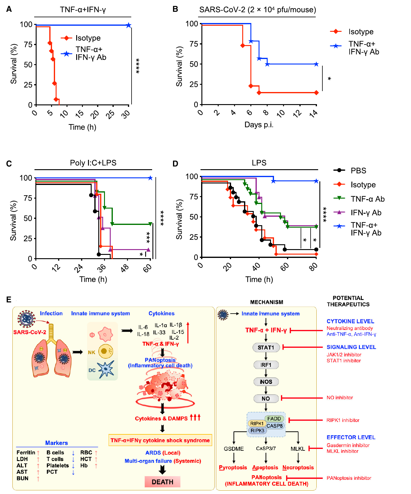
Conclusion: TNF-α + IFN-γ activates caspase-8-led PANoptosis through the STAT1/IRF1–iNOS/NO axis. This is a molecular pathway through which cytokine storm causes tissue damage and also provides a therapeutic framework with several levels at which to intervene.
III. Implications for experiments
- Exhaustive combination testing outperforms plausible guesses: Testing all 28 combinations was what excluded the more widely discussed factors IL-6 and IL-1β—when identifying key variables in a complex system, selecting candidates by intuition can miss counterintuitive answers.
- Check markers of several pathways together when identifying a mode of cell death: Measuring PI alone only establishes that "the cells died"; simultaneous assessment of GSDMD/GSDME, caspase-3/7/8, and pMLKL revealed that all three pathways were activated and GSDMD was inactivated—an incomplete set of measurements can lead to misclassification of the mode of death.
- Target an upstream convergence point when downstream pathways are redundant: Deleting any single execution molecule did not fully block death, and only deletion of caspase-8/FADD provided complete protection—for downstream pathways acting in parallel, an intervention target should be selected at their upstream point of convergence.
IV. Connections to other papers on this site
- Comparison | GSDMD is the executor of pyroptosis (He 2015, Cell Res): He established caspase-1–GSDMD as the canonical pyroptosis axis; in this paper, caspase-1 is not activated and GSDMD is instead cleaved into inactive P20, while GSDME forms the pores—the upstream caspase determines which gasdermin serves as its substrate.
- Comparison | RIPK3 cleavage limits NLRP3 (Tran 2024, Cell Death Differ): In this paper, caspase-8 drives PANoptosis; in Tran, it cleaves RIPK3 to limit pyroptosis—the same caspase-8 has effects whose direction depends on its substrate.
- Comparison | TLR-induced death in IFN-γ-primed macrophages (Simpson 2022, Immunity): This paper proposes iNOS/NO as the lethal mechanism and suggests its involvement in HLH; Simpson tested this directly in gene-knockout mice and found that HLH mortality depends on caspase-8 but not iNOS—in vitro mechanisms and their contribution in vivo need to be assessed separately.
- Extension | Caspase-8 auto-cleavage regulates the apoptosis/necroptosis switch (Li 2022, Cell Death Differ): In this paper, caspase-8/FADD drives PANoptosis; Li showed that caspase-8 auto-cleavage determines whether TNF-induced death proceeds through apoptosis or necroptosis—the processing state of caspase-8 determines the cell-death route.
(The comparisons across papers above are integrated interpretations, not direct conclusions of any single original paper.)
What remains unresolved
Limitations (two points): (1) Protection in the infection model was incomplete (antibodies were first given on day 1), so the causal chain was not fully closed; (2) how NO activates caspase-8 remains unresolved, and the mechanism was established primarily in BMDMs.
Synergism of TNF-α and IFN-γ Triggers Inflammatory Cell Death, Tissue Damage, and Mortality in SARS-CoV-2 Infection and Cytokine Shock Syndromes
Karki Rajendra; Sharma Bhesh Raj; Tuladhar Shraddha; Williams Evan Peter; Zalduondo Lillian; Samir Parimal; Zheng Min; Sundaram Balamurugan; Banoth Balaji; Malireddi R.K. Subbarao; Schreiner Patrick; Neale Geoffrey; Vogel Peter; Webby Richard; Jonsson Colleen Beth; Kanneganti Thirumala-Devi Cell. 2021;184:149–168.e17.
Figures are from the verified original paper stored in the library. Original labels, axes and controls are retained; figure numbers and sources are registered in the library.
References
- Karki R, Sharma BR, Tuladhar S, et al. Synergism of TNF-α and IFN-γ triggers inflammatory cell death, tissue damage, and mortality in SARS-CoV-2 infection and cytokine shock syndromes. Cell. 2021;184(1):149-168.e17. doi:10.1016/j.cell.2020.11.025
- Wang Y, Gao W, Shi X, et al. Chemotherapy drugs induce pyroptosis through caspase-3 cleavage of a gasdermin. Nature. 2017;547(7661):99-103.
- Christgen S, Zheng M, Kesavardhana S, et al. Identification of the PANoptosome: a molecular platform triggering pyroptosis, apoptosis, and necroptosis (PANoptosis). Front Cell Infect Microbiol. 2020;10:237.
- Selleri C, Sato T, Anderson S, Young NS, Maciejewski JP. Interferon-gamma and tumor necrosis factor-alpha suppress both early and late stages of hematopoiesis and induce programmed cell death. J Cell Physiol. 1995;165(3):538-546.
Paper discussion
No sign-up. Nickname optional.