- 01
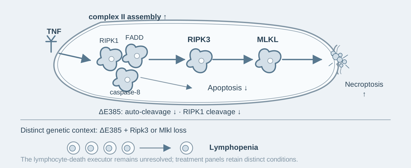
Main finding: Mice lacking the caspase-8 auto-cleavage site (ΔE385) are viable, but TNF-induced apoptosis switches to necroptosis;
- 02
Key mechanism: Loss of auto-cleavage reduces RIPK1 cleavage and enhances complex II assembly, making the RIPK1-RIPK3-MLKL pathway easier to activate;
- 03
Significance: Auto-cleavage cooperates with RIPK3/MLKL to maintain lymphocyte homeostasis, which depends on the scaffold function of RIPK1 rather than its kinase activity.
On this page
I. What question does this paper answer?
caspase-8 initiates death-receptor apoptosis and inhibits RIPK3-MLKL necroptosis (a form of programmed necrosis executed by MLKL phosphorylated by RIPK3). Its activation depends on auto-cleavage at D387, but the physiological role of this cleavage in the whole animal was unclear. The study asks: how does selectively disrupting caspase-8 auto-cleavage while retaining its other functions affect cell-death regulation and immune homeostasis in mice? The conclusions are that auto-cleavage is required for apoptosis to proceed fully; its loss switches TNF-induced apoptosis to necroptosis; and it cooperates with RIPK3/MLKL to maintain lymphocyte homeostasis, which depends on the scaffold function of RIPK1 rather than its kinase activity.
II. Following the evidence
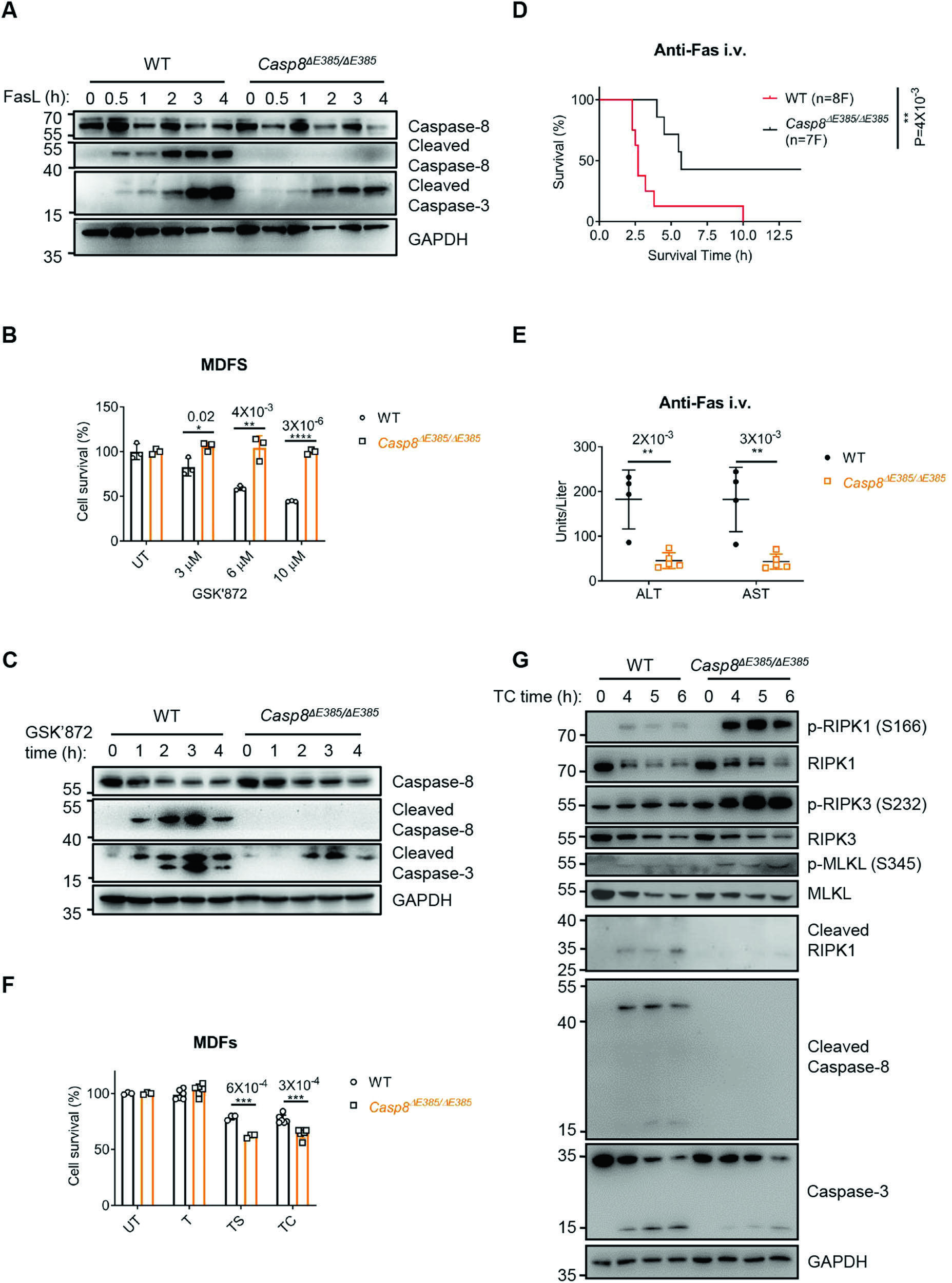
First step: mice lacking the auto-cleavage site are viable but have a mild reduction in splenic CD8+ T cells (Fig. 1). Activation of caspase-8 requires auto-cleavage at D387 between the large and small catalytic subunits. The authors deleted E385 from the recognition sequence at this site to generate Casp8ΔE385/ΔE385 knock-in mice. The mice were born and developed normally, with normal caspase-8 expression across tissues; their only changes were a mild reduction in splenic CD8+ T cells and splenomegaly. These results indicate that caspase-8 auto-cleavage is dispensable for developmental viability but has a mild effect on lymphocyte homeostasis.
Second step: loss of auto-cleavage impairs apoptosis (Fig. 2). The authors treated mouse thymocytes with the anti-Fas antibody Jo2 and administered it to mice intravenously. ΔE385 cells showed weaker apoptosis, while the mice had lower mortality and reduced liver-injury markers ALT/AST. When apoptosis was induced in mouse dermal fibroblasts (MDF) with the RIPK3 inhibitor GSK'872, ΔE385 cells were likewise markedly resistant and showed reduced caspase-3 cleavage. These results indicate that caspase-8 auto-cleavage is required for extrinsic apoptosis to proceed fully.
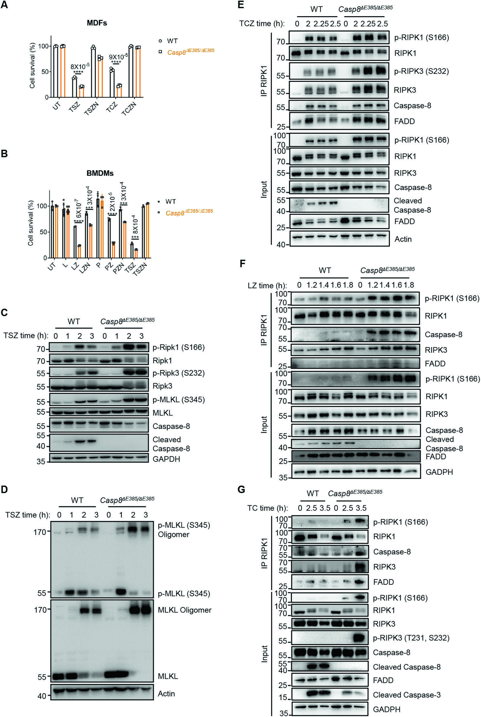
Third step: apoptosis switches to necroptosis (Figs. 2–4). The authors treated MDF with TNF-α plus a Smac mimetic (a small molecule that promotes degradation of IAP proteins). Wild-type cells underwent apoptosis, whereas death of ΔE385 cells could be rescued by Nec-1 (a RIPK1 kinase inhibitor) and was accompanied by reduced RIPK1 cleavage and enhanced assembly of complex II (a cytosolic death complex containing RIPK1, FADD, and caspase-8). In an in vivo TNF-α shock model, ΔE385 mice showed increased mortality with severe hypothermia, which was markedly alleviated on a Ripk3-knockout or kinase-inactive RIPK1 (K45A) background. These results indicate that caspase-8 lacking auto-cleavage has a reduced ability to cleave RIPK1, complex II assembles more readily, and TNF-induced apoptosis consequently switches to RIPK3-MLKL-dependent necroptosis.
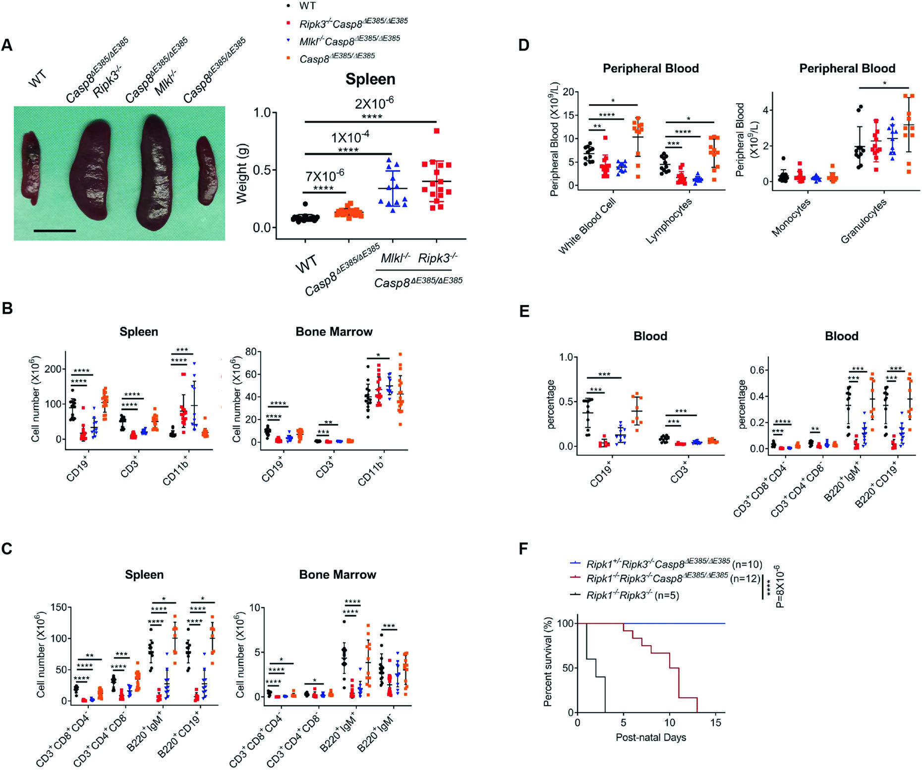
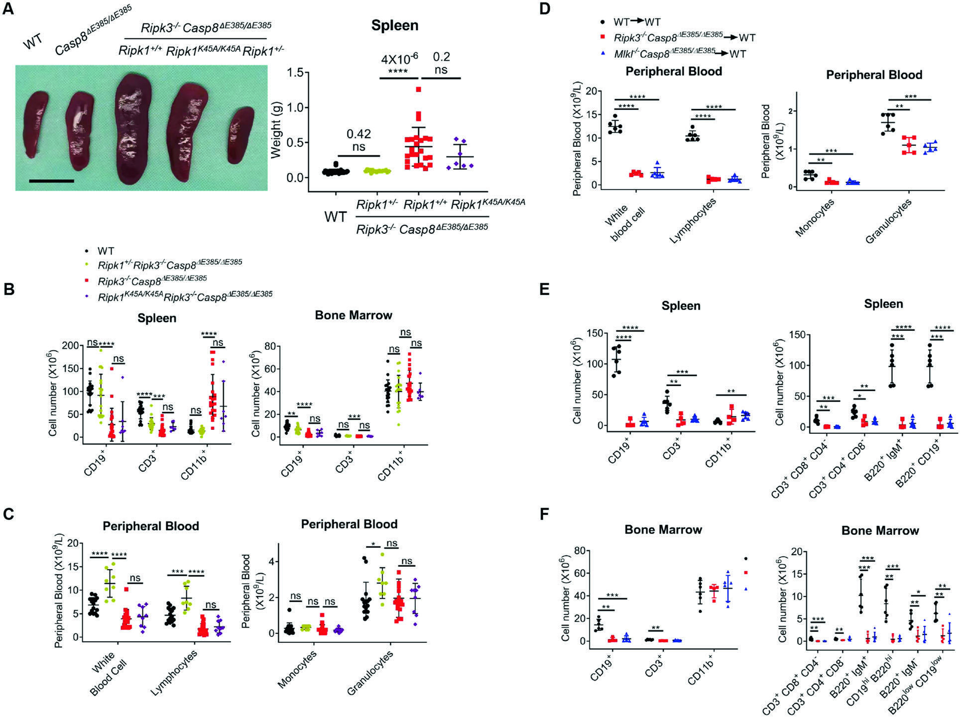
Fourth step: auto-cleavage cooperates with RIPK3/MLKL to maintain lymphocyte homeostasis (Fig. 5). Casp8−/−Ripk3−/− mice with a complete caspase-8 knockout develop lymphoproliferative disease resembling human autoimmune lymphoproliferative syndrome (ALPS). The authors found that Casp8ΔE385/ΔE385Ripk3−/− and Casp8ΔE385/ΔE385Mlkl−/− mice did not develop lymphoproliferation; instead, B- and T-cell numbers in the spleen and bone marrow fell substantially, while the proportion of myeloid cells increased. In addition, Ripk1−/−Ripk3−/−Casp8ΔE385/ΔE385 triple-mutant mice were born normally but developed skin lesions and all died at approximately 2 weeks of age, surviving longer than Ripk1−/− mice, which die in the perinatal period. These results indicate that loss of auto-cleavage and complete knockout produce opposite immune phenotypes—lymphopenia rather than lymphocyte accumulation.
Fifth step: lymphopenia is determined by the dosage of RIPK1 scaffold function (Fig. 6). The authors halved RIPK1 expression (Ripk1+/−) and found that lymphopenia and the myeloid shift in ΔE385;Ripk3−/− mice were largely reversed; kinase-inactive RIPK1 (K45A) did not reverse them. Bone-marrow transplantation demonstrated that the phenotype was intrinsic to hematopoietic cells and could be transferred with the transplant. These results indicate that maintaining lymphocyte homeostasis requires RIPK1 scaffold function rather than kinase activity and is sensitive to dosage.
III. Implications for experiments
- "Unable to auto-cleave" does not mean "caspase-8 has no function": Complete knockout is embryonically lethal, whereas cleavage-site mutants can survive to adulthood. In caspase-8 experiments, knockout, catalytically inactive mutations (C362A/S), and cleavage-site mutations (D387A/ΔE385) have profoundly different phenotypes; define the question before choosing the genotype.
- More cell death does not mean stronger apoptosis: ΔE385 cells actually show more death after TNF+Smac treatment, so measuring only the death rate will lead to a mistaken conclusion; Nec-1 rescue and pRIPK1/pRIPK3/pMLKL blots need to be considered together to confirm that the mode of death has switched.
- In vivo TNF shock models require accompanying body-temperature monitoring: Survival data together with severe hypothermia suggest a necroptotic component, which can be checked against a Ripk3-knockout or kinase-inactive RIPK1 background.
IV. Related papers on this site
- Extension | TNF-α+IFN-γ synergistic PANoptosis (Karki 2021, Cell): In Karki, caspase-8/FADD drive PANoptosis; this paper shows that caspase-8 auto-cleavage determines whether TNF-induced death follows apoptosis or necroptosis—the processing state of caspase-8 determines the death route.
- Extension | MLKL membrane trafficking controls necroptosis kinetics (Samson 2020, Nat Commun): This paper shows that loss of auto-cleavage redirects death toward necroptosis; Samson describes the downstream trafficking and threshold of MLKL from phosphorylation to membrane rupture—respectively, "which route to take" and "how to follow that route".
- Comparison | Effector caspases amplify apoptotic signals through feedback (McComb 2019, Sci Adv): Both studies show reduced apoptotic efficiency, but at different mechanistic points. Here, loss of auto-cleavage in the initiator caspase-8 weakens apoptosis; in McComb, loss of the effector caspase-3/-7 attenuates upstream signals—there are amplification mechanisms at both ends of the apoptotic cascade.
(The cross-paper comparisons above are a synthesis, not a direct conclusion of any individual original paper.)
What remains unresolved
Limitations: ① The spectrum of effects of residual ΔE385 enzymatic activity on different substrates was not systematically measured; ② the specific death executor underlying lymphopenia in ΔE385;Ripk3−/− mice was not fully defined.
Caspase-8 auto-cleavage regulates programmed cell death and collaborates with RIPK3/MLKL to prevent lymphopenia
Xiaoming Li; Fang Li; Xixi Zhang; Haiwei Zhang; Qun Zhao; Ming Li; Xiaoxia Wu; Lingxia Wang; Jianling Liu; Xuanhui Wu; Yangjing Ou; Mingyan Xing; Yue Zhang; Jiangshan Deng; Xiuzhe Wang; Yan Luo; Jinbao Li; Yuwu Zhao; Haibing Zhang Cell Death & Differentiation. 2022;29:1500–1512.
Figures are from the verified original paper stored in the library. Original labels, axes and controls are retained; figure numbers and sources are registered in the library.
References
- Li X, Li F, Zhang X, et al. Caspase-8 auto-cleavage regulates programmed cell death and collaborates with RIPK3/MLKL to prevent lymphopenia. Cell Death Differ. 2022;29(8):1500-1512. doi:10.1038/s41418-022-00938-9
- Newton K, Wickliffe KE, Dugger DL, et al. Cleavage of RIPK1 by caspase-8 is crucial for limiting apoptosis and necroptosis. Nature. 2019;574(7778):428-431. doi:10.1038/s41586-019-1270-x
- Kaiser WJ, Upton JW, Long AB, et al. RIP3 mediates the embryonic lethality of caspase-8-deficient mice. Nature. 2011;471(7338):368-372. doi:10.1038/nature09857
- Alvarez-Diaz S, Dillon CP, Lalaoui N, et al. The pseudokinase MLKL and the kinase RIPK3 have distinct roles in autoimmune disease caused by loss of death-receptor-induced apoptosis. Immunity. 2016;45(3):513-526. doi:10.1016/j.immuni.2016.07.016
Paper discussion
No sign-up. Nickname optional.