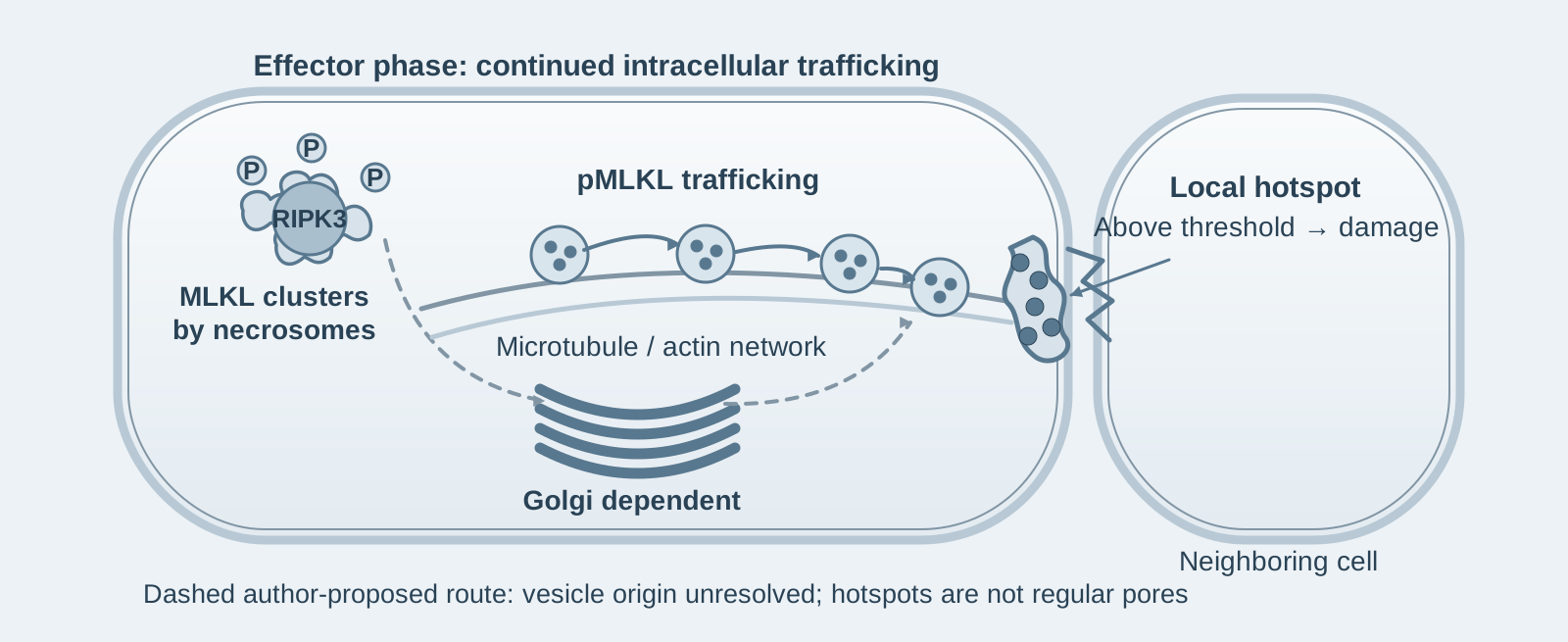
- 01
Main finding: A substantial delay separates MLKL phosphorylation from membrane lysis. Phosphorylated MLKL is transported in vesicles and accumulates at 1–2 hotspots on the plasma membrane; rupture occurs only after a threshold is exceeded;
- 02
Key mechanism: Trafficking depends on the Golgi–microtubule–actin network and shares a route with tight junction proteins, including ZO-1; hotspots preferentially occur at intercellular junctions;
- 03
Research significance: Necroptosis has two further checkpoints downstream of the necrosome—trafficking, which determines its speed, and a membrane threshold, which determines the extent of lysis—providing new targets for intervention.
On this page
1. What this paper answers
In necroptosis, RIPK3-mediated phosphorylation of MLKL is considered a key step in executing cell death, but what happens between phosphorylation and membrane rupture remains unclear. This paper asks: What determines this delay, and how does MLKL reach the plasma membrane and cause lysis? It concludes that phosphorylated MLKL first assembles into cytoplasmic clusters and is then transported in vesicles through Golgi, microtubule and actin networks to the plasma membrane, forming 1–2 hotspots at intercellular junctions. Membrane rupture occurs only when MLKL accumulation at these hotspots exceeds a threshold; lysis starts at the hotspots and can be passed to adjacent cells. Trafficking speed and the membrane threshold therefore constitute two new checkpoints downstream of the necrosome.
2. The chain of evidence
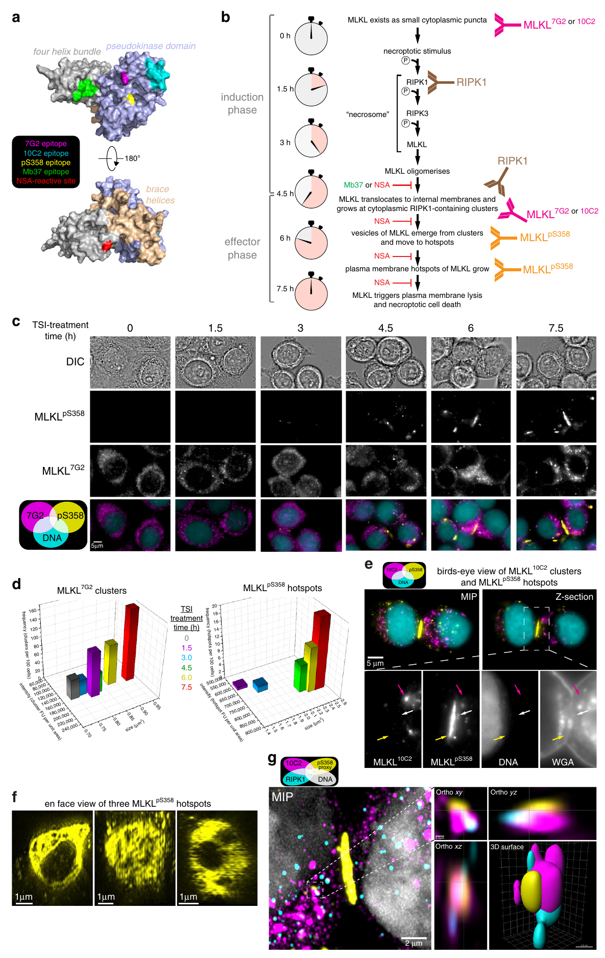
Step 1: Confirming the delay between phosphorylation and death and establishing tools for single-cell tracking (Figures 1–2). The authors induced necroptosis in HT29 cells with TSI (TNF + a Smac mimetic + the caspase inhibitor IDN-6556). pS358-MLKL appeared at 3 hours and peaked at 6 hours, while LDH release lagged by more than 3 hours. The authors generated the 7G2 and 10C2 monoclonal antibodies, which recognize different epitopes, and combined them with a pS358 phosphorylation-specific antibody to track endogenous MLKL by immunofluorescence. At rest, MLKL appeared as small cytoplasmic puncta; during the effector phase, it assembled into large clusters around the necrosome—a high-molecular-weight complex assembled from RIPK1 and RIPK3. Phosphorylated MLKL subsequently appeared at the plasma membrane in vesicles. These results indicate that the delay occurs after MLKL membrane association.
Step 2: MLKL forms hotspots on the plasma membrane, and rupture requires a threshold to be exceeded (Figures 2–3). The authors found that phosphorylated MLKL was distributed unevenly on the plasma membrane, forming 1–2 hotspots per cell, approximately 0.5–3 μm in diameter. These hotspots preferentially occurred at intercellular junctions and had an irregular ring-like shape and heterogeneous internal structure—a morphology that does not support a model in which MLKL assembles regular membrane pores. When MLKL membrane translocation was blocked by the synthetic protein ligand Mb37, which binds the MLKL killer domain, or by the compound NSA, both hotspots and death were inhibited. These results indicate that necroptosis depends on plasma membrane MLKL locally exceeding a threshold, rather than being uniformly distributed across the entire membrane.
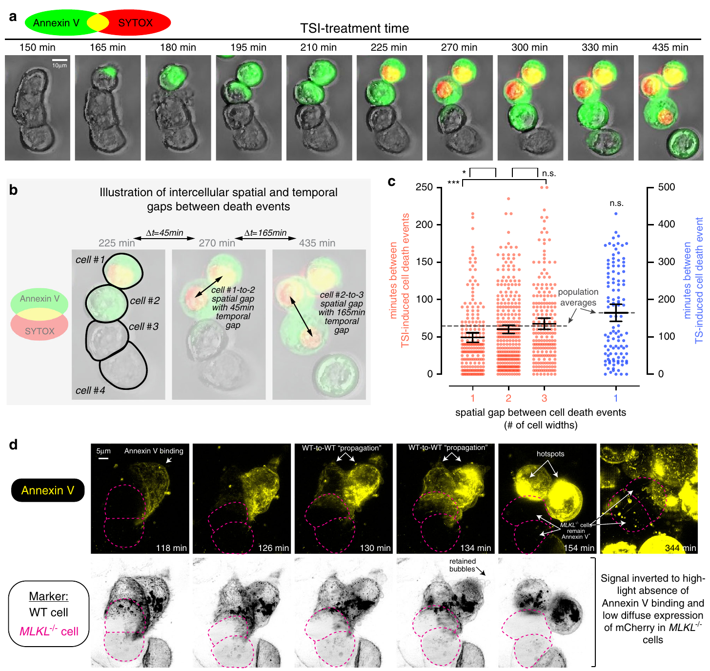
Step 3: Hotspots are the starting points of membrane damage, which can be passed to adjacent cells (Figures 4–5). Using real-time live-cell imaging with lattice light sheet microscopy, the authors found that membrane damage, indicated by Annexin V binding, started precisely at the hotspots. Statistics from a large sample showed that cells adjacent to dying cells underwent necroptosis approximately 25% faster than cells that were not adjacent; this neighborhood effect was absent in TNF-induced apoptosis. These results indicate that membrane damage at hotspots can spread to neighboring cells along intercellular junctions, so necroptosis in epithelial cells has an effect that promotes death between cells.
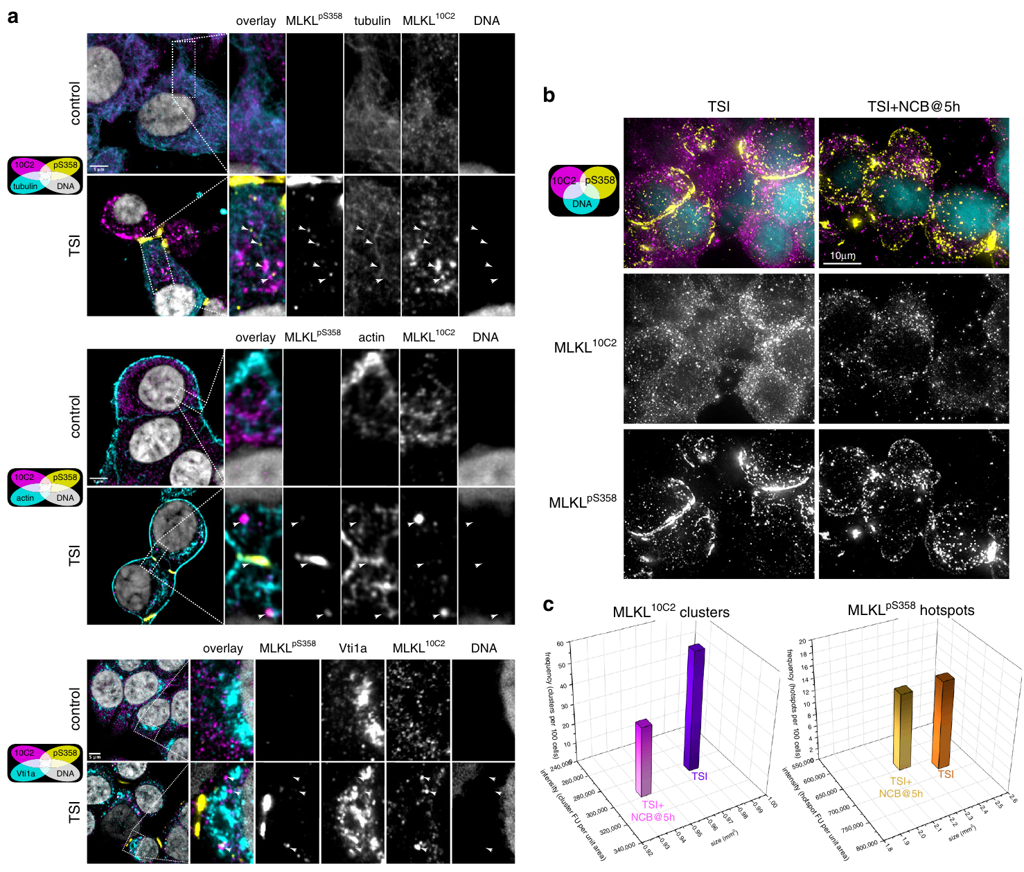
Step 4: Intracellular trafficking determines death kinetics (Figures 6–7). The authors used Nocodazole, Cytochalasin B and Brefeldin A to disrupt the microtubule, actin and Golgi networks, respectively, and found that the three agents combined as NCB inhibited membrane lysis. Crucially, adding NCB or NSA after 5 hours of TSI treatment—when phosphorylated MLKL had already begun to reach the membrane—still blocked death in three human epithelial cell lines: HT29, HCC2998 and SW620. These results indicate that MLKL trafficking to the plasma membrane continues throughout the effector phase and remains necessary throughout it, making this a step downstream of the necrosome that can be targeted pharmacologically.
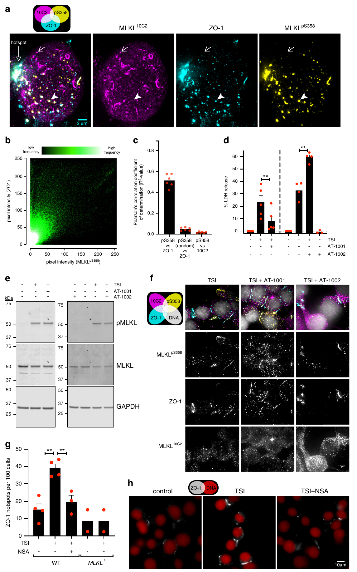
Step 5: MLKL shares a trafficking route with tight junction proteins (Figure 8). The authors co-stained phosphorylated MLKL with various cellular markers and found substantial colocalization with the tight junction proteins ZO-1, occludin and JAM-A, with quantitative correlation at the pixel level. They found no colocalization with markers of desmosomes, adherens junctions, endosomes, mitochondria or lysosomes. AT-1001, which stabilizes the organization of ZO-1 junctions, opposed necroptosis, whereas AT-1002, which disrupts junctions, promoted it; neither affected the level of MLKL phosphorylation. These results indicate that MLKL and tight junction proteins travel via the same route, and the organization of intercellular junctions directly determines the threshold for membrane rupture.
3. Implications for experiments
- Use "delayed drug addition" to distinguish the phase in which an inhibitor acts: If adding a drug 5 hours after TSI still blocks death, its action occurs after MLKL phosphorylation. Adding drugs separately during the induction and effector phases is a straightforward way to locate the step at which they act.
- Stage MLKL immunofluorescence by morphology: Small cytoplasmic puncta → large clusters beside necrosomes → plasma membrane hotspots correspond to three steps. A pS358 Western blot alone cannot establish how far trafficking has progressed.
- Necroptosis in epithelial cells has a neighborhood effect: Seeding density and the state of intercellular junctions affect death kinetics; confluence should be kept consistent when comparing experiments.
4. Related papers on this site
- Further reading | Active MLKL activates NLRP3 in a cell-intrinsic manner (Conos 2017, PNAS): Conos showed that active MLKL activates NLRP3 through potassium efflux before cell rupture. This paper defines the trafficking of phosphorylated MLKL from cytoplasmic clusters to plasma membrane hotspots and the threshold for membrane rupture. The "before rupture" window in Conos is precisely the trafficking and accumulation period characterized here.
- Further reading | Caspase-8 self-cleavage regulates the switch between apoptosis and necroptosis (Li 2022, Cell Death Differ): Li showed that loss of caspase-8 self-cleavage switches TNF-induced apoptosis to necroptosis. This paper supplies details of MLKL trafficking and membrane lysis kinetics at the execution stage after that switch.
(These comparisons across papers are an integrated interpretation, rather than a direct conclusion of any individual source paper.)
What remains unresolved
Limitations: ① All conclusions come from epithelial cell lines; whether hotspots and co-trafficking are widespread in other cell types has not been tested. ② The vesicle types and adaptor molecules that carry MLKL and tight junction proteins have not been identified.
MLKL trafficking and accumulation at the plasma membrane control the kinetics and threshold for necroptosis
Andre L. Samson; Ying Zhang; Niall D. Geoghegan; Xavier J. Gavin; Katherine A. Davies; Michael J. Mlodzianoski; Lachlan W. Whitehead; Daniel Frank; Sarah E. Garnish; Cheree Fitzgibbon; Anne Hempel; Samuel N. Young; Annette V. Jacobsen; Wayne Cawthorne; Emma J. Petrie; Maree C. Faux; Kristy Shield-Artin; Najoua Lalaoui; Joanne M. Hildebrand; John Silke; Kelly L. Rogers; Guillaume Lessene; Edwin D. Hawkins; James M. Murphy Nature Communications. 2020;11:3151.
Figures are from the verified original paper stored in the library. Original labels, axes and controls are retained; figure numbers and sources are registered in the library.
References
- Samson AL, Zhang Y, Geoghegan ND, et al. MLKL trafficking and accumulation at the plasma membrane control the kinetics and threshold for necroptosis. Nat Commun. 2020;11:3151. doi:10.1038/s41467-020-16887-1
- Conos SA, Chen KW, De Nardo D, et al. Active MLKL triggers the NLRP3 inflammasome in a cell-intrinsic manner. Proc Natl Acad Sci USA. 2017;114(6):E961-E969. doi:10.1073/pnas.1613305114
- Murphy JM, Czabotar PE, Hildebrand JM, et al. The pseudokinase MLKL mediates necroptosis via a molecular switch mechanism. Immunity. 2013;39(3):443-453. doi:10.1016/j.immuni.2013.06.018
Paper discussion
No sign-up. Nickname optional.