- 01
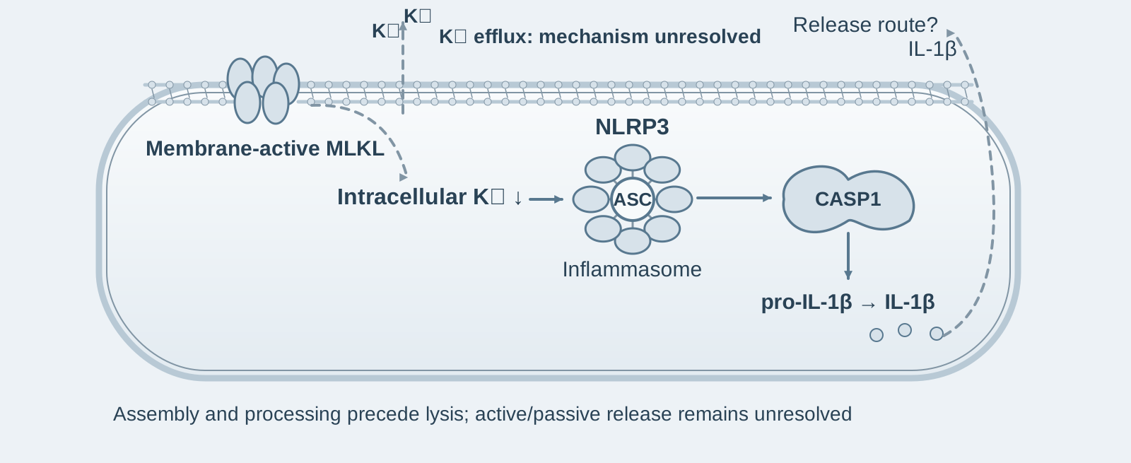
Main finding: During necroptosis, MLKL activates NLRP3–caspase-1 before cell lysis, driving IL-1β processing and secretion; this process is cell-intrinsic;
- 02
Key mechanism: This requires the 4HB killing domain, oligomerization, and membrane binding of MLKL, and activates NLRP3 through a decrease in intracellular potassium concentration; GSDMD is not an essential release channel;
- 03
Significance: The inflammatory potential of necroptosis primarily comes from NLRP3–IL-1β rather than passive DAMP release, suggesting that NLRP3 inhibitors could be used in MLKL-driven diseases.
On this page
1. What question does this paper answer?
Why does necroptosis cause inflammation? The prevailing view is that cell rupture releases DAMPs that activate surrounding cells. This paper asks: Is MLKL activation of NLRP3 a cell-intrinsic event or a secondary event after rupture? How much does it contribute to inflammation? The conclusion is that the membrane-damaging activity of MLKL lowers intracellular potassium and activates NLRP3–caspase-1 before cell rupture, completing IL-1β processing and secretion; experiments transferring supernatant from dying cells to unstimulated cells demonstrate that this active pathway accounts for most of the inflammatory potential of necroptosis, rather than DAMPs leaking out after rupture.
2. The logic of the evidence
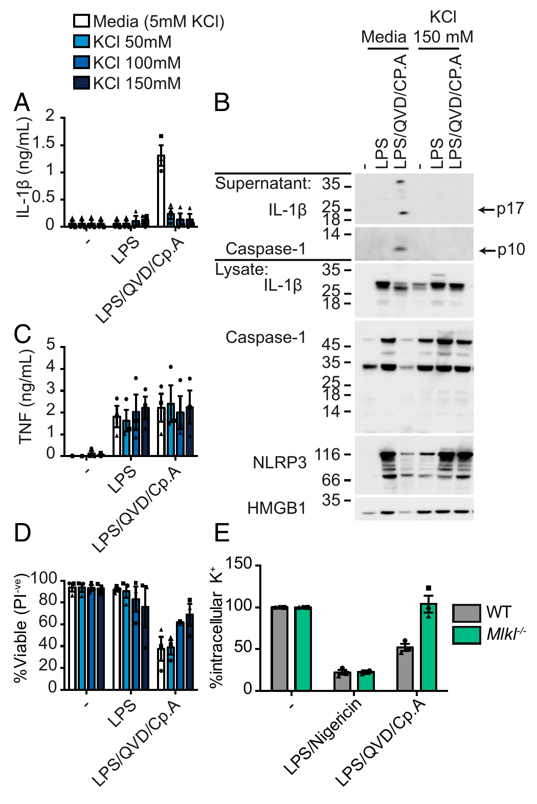
Step 1: MLKL activates NLRP3 through potassium efflux (Fig. 1). The authors induced necroptosis in mouse bone marrow-derived macrophages (BMDMs) using LPS + QVD (a pan-caspase inhibitor) + Cp.A (a Smac mimetic). Mass spectrometry showed a decrease in intracellular potassium concentration, whereas no such decrease occurred in Mlkl-knockout cells; adding 50–150 mM potassium chloride extracellularly inhibited IL-1β secretion in a dose-dependent manner without affecting TNF secretion. Genetically, knockout of Nlrp3 or caspase-1 blocked IL-1β secretion but did not block necroptotic death. Together, these results indicate that MLKL activates NLRP3 at the level of death execution through potassium efflux; NLRP3 controls inflammation but not death.
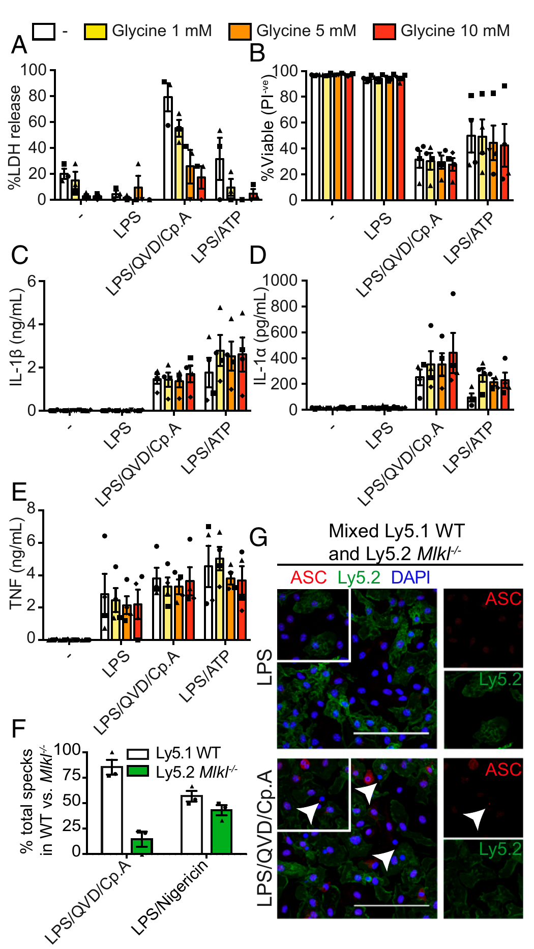
Step 2: NLRP3 activation is a cell-intrinsic event (Figs. 3–4). MLKL-induced IL-1β secretion was normal in GSDMD-knockout BMDMs, indicating that release through this pathway does not require GSDMD pores. The authors treated cells with glycine (an osmoprotectant that delays the swelling and rupture of cells undergoing lysis) and found that release of large molecules such as LDH was suppressed, while IL-1β processing and secretion were unaffected; imaging of mixed cultures of WT and Mlkl-knockout cells showed that ASC specks formed only within wild-type cells. Together, these results indicate that NLRP3 activation occurs before cell rupture and is completed within the dying cell, rather than being a secondary event in which DAMPs stimulate neighboring cells.
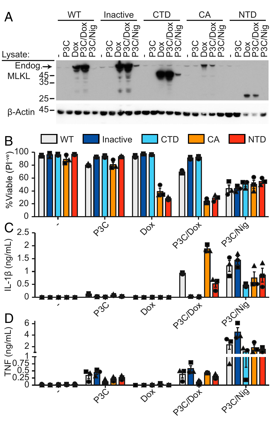
Step 3: Only MLKL capable of damaging membranes activates NLRP3 (Figs. 5–6). The authors used doxycycline to induce expression of a series of MLKL constructs in THP1 cells: wild-type MLKL, a constitutively active mutant (S345D), and a fragment containing only the N-terminal 4HB killing domain (1–180) all killed cells and induced IL-1β secretion; a membrane-binding-defective mutant (E109A/E110A) and a C-terminal fragment lacking the 4HB (124–464) neither killed cells nor induced IL-1β. The NLRP3 inhibitor MCC950 blocked IL-1β secretion without affecting cell death or TNF. Together, these results indicate that the ability of MLKL to activate NLRP3 is linked to its membrane-damaging activity—the two share the same structural basis (the 4HB domain and membrane binding).
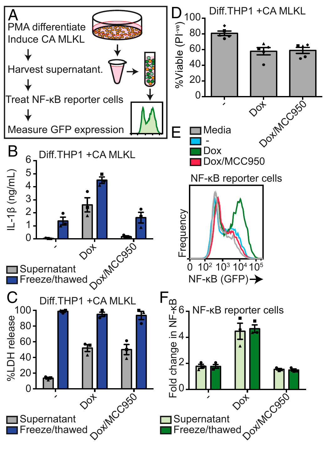
Step 4: Supernatant transfer experiments demonstrate that inflammation comes from NLRP3 rather than DAMPs (Fig. 7). The authors killed PMA-differentiated THP1 cells with constitutively active MLKL (PMA priming avoids contamination of the supernatant with TLR ligands), transferred the supernatant to naïve THP1 cells bearing an NF-κB reporter gene, and found that NF-κB was activated; however, if the dying cells had first been pretreated with MCC950 (with death still occurring as usual), the supernatant lost its ability to activate the reporter cells. Conversely, supernatant from cells completely lysed by freeze-thawing, releasing all their contents (including pro-IL-1β), also failed to activate NF-κB. Together, these results indicate that inflammatory signals from necroptotic cells mainly come from mature IL-1β generated by NLRP3, while contents released by cell rupture make only a small contribution.
3. Implications for experiments
- MCC950 is a tool for separating "death" from "inflammation": it blocks IL-1β without blocking necroptotic death—it can be used to test whether a phenotype is driven by death itself or by NLRP3-mediated inflammation.
- Glycine + high potassium + gene knockout allow "lysis" and "secretion" to be tested separately; however, note the limitation stated by the authors themselves: glycine only delays membrane rupture rather than completely blocking it, so PI positivity must not be equated with complete rupture when interpreting the results.
- GSDMD is not the default channel for every release event downstream of caspase-1: during necroptosis, secretion after caspase-1 processing of IL-1β does not depend on GSDMD—when designing experiments on "release mechanisms," do not apply Gsdmd knockout as a one-size-fits-all approach.
4. Related papers on this site
- Extension | MLKL membrane trafficking controls the kinetics of necroptosis (Samson 2020, Nat Commun): This paper demonstrates that active MLKL activates NLRP3 through potassium efflux before cell rupture; Samson defines the trafficking of phosphorylated MLKL from cytoplasmic clusters to plasma membrane hotspots and the threshold for membrane rupture—the "before rupture" time window in this paper is precisely the trafficking and accumulation period characterized by Samson.
- Comparison | Apoptosis activates NLRP3 through pannexin-1 (Chen 2019, EMBO J): This paper demonstrates that, during necroptosis, the membrane activity of MLKL activates NLRP3 through potassium efflux before rupture; Chen demonstrates that the pannexin-1 channel serves the same role during apoptosis—different modes of cell death share the logic of "potassium efflux → NLRP3," but use different channels.
(The cross-paper comparisons above are a synthesis, not direct conclusions of either original paper.)
What remains unresolved
Limitations: ① Whether IL-1β is actively secreted or passively leaks out during necroptosis remains unresolved; ② the molecular details of how MLKL causes potassium efflux (direct pore formation or action through ion channels) were not defined; ③ its contribution in in vivo disease models is an indirect inference.
Active MLKL triggers the NLRP3 inflammasome in a cell-intrinsic manner
Stephanie A. Conos; Kaiwen W. Chen; Dominic De Nardo; Hideki Hara; Lachlan Whitehead; Gabriel Núñez; Seth L. Masters; James M. Murphy; Kate Schroder; David L. Vaux; Kate E. Lawlor; Lisa M. Lindqvist; James E. Vince Proceedings of the National Academy of Sciences of the United States of America. 2017;114:E961-E969.
Figures are from the verified original paper stored in the library. Original labels, axes and controls are retained; figure numbers and sources are registered in the library.
References
- Conos SA, Chen KW, De Nardo D, et al. Active MLKL triggers the NLRP3 inflammasome in a cell-intrinsic manner. Proc Natl Acad Sci USA. 2017;114(6):E961-E969. doi:10.1073/pnas.1613305114
- Gutierrez KD, Davis MA, Daniels BP, et al. MLKL activation triggers NLRP3-mediated processing and release of IL-1β independently of gasdermin-D. J Immunol. 2017;198(5):2156-2164. doi:10.4049/jimmunol.1601757
- Lawlor KE, Khan N, Mildenhall A, et al. RIPK3 promotes cell death and NLRP3 inflammasome activation in the absence of MLKL. Nat Commun. 2015;6:6282. doi:10.1038/ncomms7282
Paper discussion
No sign-up. Nickname optional.