- 01
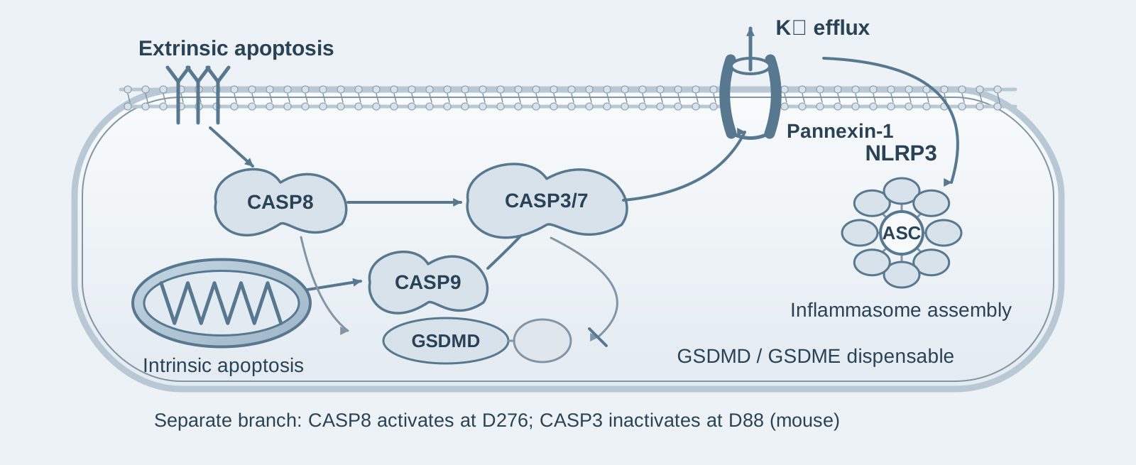
Main finding: During extrinsic apoptosis, caspase-8 directly cleaves GSDMD to cause cell lysis, in parallel with caspase-3/7-dependent secondary necrosis;
- 02
Key mechanism: Caspase-3 cleaves and inactivates GSDMD at D88, restricting cell lysis; NLRP3 activation is driven by pannexin-1 rather than gasdermin pores;
- 03
Significance: Pannexin-1 is the common channel through which intrinsic and extrinsic apoptosis activate NLRP3; the "immunological silence" of apoptosis is actively maintained.
On this page
I. What question does this paper answer?
Caspase-8/-9-dependent apoptosis induced by drugs such as Smac mimetics, TAK1 inhibitors and BH3 mimetics causes cell lysis and activates the NLRP3 inflammasome. This contradicts the classical view of "immunologically silent apoptosis", and the mechanism is unclear. The paper concludes that during extrinsic apoptosis, caspase-8 directly cleaves GSDMD to drive cell lysis, which is normally restricted by inactivating cleavage at D88 by caspase-3. Cell lysis during intrinsic apoptosis, by contrast, is independent of gasdermins. Both forms of apoptosis activate the pannexin-1 channel through caspase-3/7, and potassium efflux then activates NLRP3.
II. How the evidence builds the argument
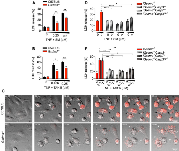
First step: GSDMD and caspase-3/7-dependent secondary necrosis act in parallel to cause cell lysis during extrinsic apoptosis (Figure 1). The authors treated macrophages with TNF plus a Smac mimetic (AZD 5582) or a TAK1 inhibitor to assemble the ripoptosome and activate caspase-8. Wild-type cells underwent lysis and displayed pyroptotic morphology (plasma membrane ballooning and PI uptake), whereas Gsdmd-knockout cells retained membrane integrity and displayed classical apoptotic morphology, although the apoptotic bodies still took up PI over time. Further CRISPR deletion of caspase-3 or caspase-7 in the Gsdmd-knockout background completely abolished cell lysis. These results indicate that membrane rupture during extrinsic apoptosis has two parallel sources: GSDMD pores and caspase-3/7-driven secondary necrosis.
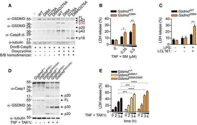
Second step: Caspase-8 and caspase-1 activate GSDMD at the same site, while caspase-3 inactivates it at another site (Figures 2–3). The authors found that GSDMD was still processed into active p30 in caspase-1/11-knockout cells. In HEK293T cells, which naturally lack caspase-1, drug-induced dimerization of caspase-8 cleaved wild-type GSDMD to produce p30, together with the p43 and p20 fragments generated by inactivating cleavage at D88. The D276A mutant accumulated only p43, while the D88A mutant accumulated only p30. An in vitro fluorescent substrate assay showed that cleavage by caspase-8 was 30-fold less efficient than cleavage by caspase-1. These results indicate that caspase-8 directly activates GSDMD at D276, whereas caspase-3 cleaves and inactivates it at D88.
Third step: D88A knock-in mice demonstrate that inactivating cleavage has a restraining role in vivo (Figure 3). The authors generated Gsdmd D88A/D88A knock-in mice, which were born at Mendelian ratios, were healthy and had normal immune systems. In their macrophages treated with TNF plus a TAK1 inhibitor or a Smac mimetic, p30 accumulated, the inactive fragments disappeared and cell death increased 1.5-fold, while caspase-1 processing remained unchanged. These results indicate that inactivating cleavage of GSDMD by caspase-3 restricts proinflammatory cell lysis driven by caspase-8, and GSDMD pores do not promote NLRP3 activation in this model.
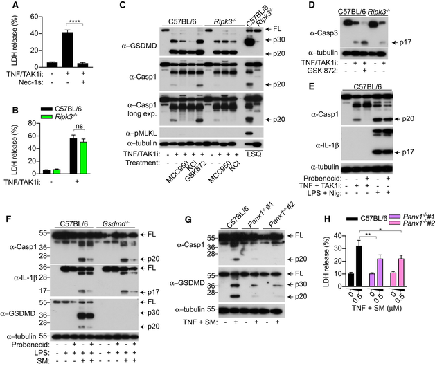
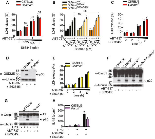
Fourth step: Both forms of apoptosis activate NLRP3 through pannexin-1, independently of gasdermins (Figures 4–5). The authors found that during extrinsic apoptosis, single or double knockout of Gsdmd and Gsdme did not affect production of caspase-1 p20, and high potassium inhibited wild-type and knockout cells equally. RIPK3 promoted caspase-3 activation through its scaffolding function, independently of kinase activity and MLKL. The pannexin-1 inhibitors probenecid and trovafloxacin markedly reduced caspase-1 activation, without affecting responses to nigericin or poly(dA:dT); Panx1 knockout likewise substantially reduced it. In the intrinsic apoptosis model (ABT-737 plus the MCL-1 inhibitor S63845), cell lysis was independent of GSDMD and GSDME, but caspase-1 processing still depended on pannexin-1. These results indicate that the pannexin-1 channel is a common route by which intrinsic and extrinsic apoptosis activate NLRP3, with gasdermin pores participating in neither case.
III. Implications for experiments
- Detecting GSDMD p30 in an apoptosis model does not by itself establish "pyroptosis": Caspase-8 activates GSDMD at only approximately 1/30 the efficiency of caspase-1, and p30 is also cleaved into p20 by caspase-3. The p30, p43 and p20 bands need to be considered together; p43/p20 represent inactivating cleavage.
- D88A knock-in mice provide a clean tool for eliminating inactivating cleavage: The animals are healthy and have normal immune systems, and can be used to test the contribution of inactivating cleavage at D88 in any apoptosis model.
- Check pannexin-1 first when attributing NLRP3 activation in an apoptotic setting: If gasdermin knockout does not alter caspase-1 activation, pannexin-1 is a candidate. Note that probenecid and trovafloxacin have limited specificity, so genetic knockout evidence is needed.
IV. Connections to other papers on this site
- Comparison | Redundant apoptotic and pyroptotic machinery in XIAP deficiency (Hughes 2023, EMBO J): The two papers agree that caspase-8 cleaves GSDMD in the same IAP-inhibition model, but reach opposite conclusions about whether NLRP3 activation downstream of extrinsic apoptosis depends on pannexin-1 (required in this paper, dispensable in Hughes). The general applicability of this channel therefore remains to be tested.
- Extension | Bidirectional crosstalk between pyroptosis and apoptosis (Taabazuing 2017, Cell Chem Biol): Taabazuing found that the apoptotic caspases caspase-3/7 cleave GSDMD into inactive fragments at D87. This paper uses D88A knock-in mice to demonstrate that this inactivating cleavage is a physiological mechanism that restricts proinflammatory cell lysis during apoptosis, advancing a phenomenological observation to in vivo evidence.
- Comparison | Active MLKL activates NLRP3 in a cell-intrinsic manner (Conos 2017, PNAS): Conos demonstrated that during necroptosis, the membrane activity of MLKL activates NLRP3 through potassium efflux before cell rupture. This paper demonstrates that pannexin-1 fulfills the same role during apoptosis. Different forms of cell death share the logic of "potassium efflux → NLRP3", but use different channels.
(The cross-paper comparisons above are an integrated interpretation, not a direct conclusion of any single original paper.)
What remains unresolved
Limitations: ① Only in vitro macrophage experiments were performed with the D88A mice; the in vivo significance of inactivating cleavage was not validated. ② The conclusions concerning pannexin-1 rely in part on inhibitors with limited specificity.
Extrinsic and intrinsic apoptosis activate pannexin-1 to drive NLRP3 inflammasome assembly
Kaiwen W Chen; Benjamin Demarco; Rosalie Heilig; Kateryna Shkarina; Andreas Boettcher; Christopher J Farady; Pawel Pelczar; Petr Broz The EMBO Journal. 2019;38:.
Figures are from the verified original paper stored in the library. Original labels, axes and controls are retained; figure numbers and sources are registered in the library.
References
- Chen KW, Demarco B, Heilig R, et al. Extrinsic and intrinsic apoptosis activate pannexin-1 to drive NLRP3 inflammasome assembly. EMBO J. 2019;38(10):e101638. doi:10.15252/embj.2019101638
- Taabazuing CY, Okondo MC, Bachovchin DA. Pyroptosis and apoptosis pathways engage in bidirectional crosstalk in monocytes and macrophages. Cell Chem Biol. 2017;24(4):507-514.e4. doi:10.1016/j.chembiol.2017.03.009
- Vince JE, De Nardo D, Gao W, et al. The mitochondrial apoptotic effectors BAX/BAK activate caspase-3 and -7 to trigger NLRP3 inflammasome and caspase-8 driven IL-1β activation. Cell Rep. 2018;25(9):2339-2353.e4. doi:10.1016/j.celrep.2018.10.103
Paper discussion
No sign-up. Nickname optional.