- 01
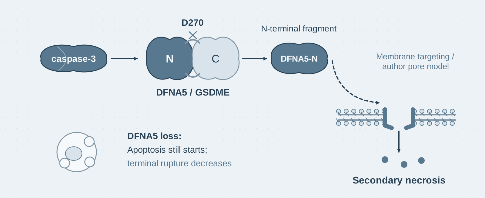
Main finding: Caspase-3 cleaves DFNA5 (GSDME) at D270, and its N-terminal fragment executes necrotic membrane rupture—secondary necrosis is a programmed event;
- 02
Key mechanism: DFNA5 shares the architecture of GSDMD: caspase-3 relieves C-terminal autoinhibition, releasing an N-terminal fragment with a membrane-targeting domain that forms pores from the inner side of the membrane;
- 03
Significance: The findings unify the terminal morphology of apoptosis and pyroptosis and explain why apoptotic cells release inflammatory contents when they are not cleared.
On this page
1. What question does this paper answer?
If apoptotic cells are not cleared promptly, they eventually swell, lose membrane integrity, and release their contents: this is secondary necrosis. It looks almost identical to pyroptosis, yet was long regarded as passive osmotic disintegration. The authors began with an unexpected observation: casp-1/11 double-knockout macrophages lack inflammatory caspases and therefore GSDMD-mediated pyroptosis, but still swell and rupture after VSV infection, releasing large amounts of LDH. Necroptosis inhibitors could not block this response, whereas the pan-caspase inhibitor zVAD could. Which caspase cleaves which substrate to cause this pyroptosis-like death without GSDMD?
2. Following the chain of evidence
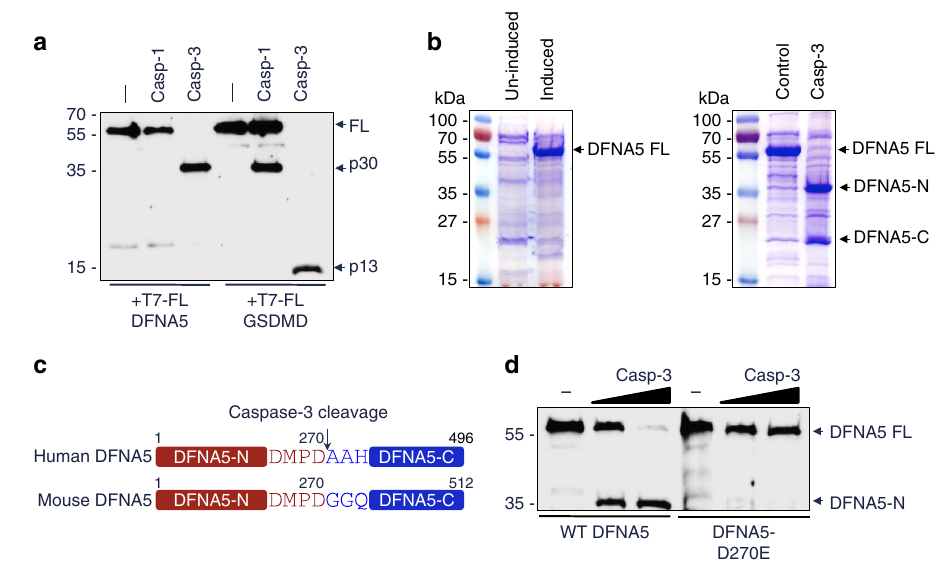
Step 1: Identify the substrate (Figs. 1–2). The candidate came from a clue in human genetics: deafness-causing mutations in DFNA5 truncate the protein near its N-terminal region, suggesting that this region might be cytotoxic, like GSDMD-N. The authors treated DFNA5 with all human caspases one by one in vitro and found that only caspase-3 cleaved it efficiently, whereas caspase-1 did not. The D270E mutation completely resisted cleavage. They then activated endogenous apoptosomes by adding cytochrome c to S100 lysates and again found cleavage of wild-type DFNA5 but not D270E. Cleavage was absent in caspase-3-deficient MCF7 cells and was restored by re-expressing caspase-3. Together, these results indicate that DFNA5 is a physiological substrate downstream of the apoptotic pathway.
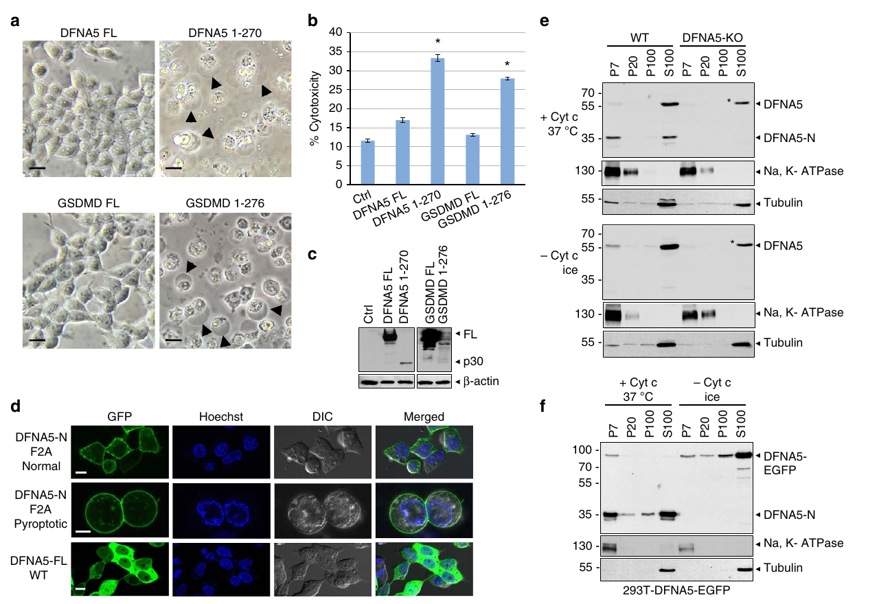
Step 2: Show that the N-terminal fragment is cytotoxic and targets the plasma membrane (Figs. 3–4). Expressing the DFNA5-N fragment in cells was sufficient to make them swell, rupture, and die; the full-length protein did not do so. zVAD could not block this effect, indicating that the fragment is the cytotoxic executor and does not require other caspases. Using the F2A mutant, whose toxicity is reduced, the authors observed DFNA5-N at the plasma membrane by microscopy, whereas the full-length protein was distributed in the cytoplasm. Homology modelling indicated that N-terminal residues 1–56 form a membrane-targeting domain. After its basic region was mutated, the fragment could neither localize to the membrane nor kill the cells.
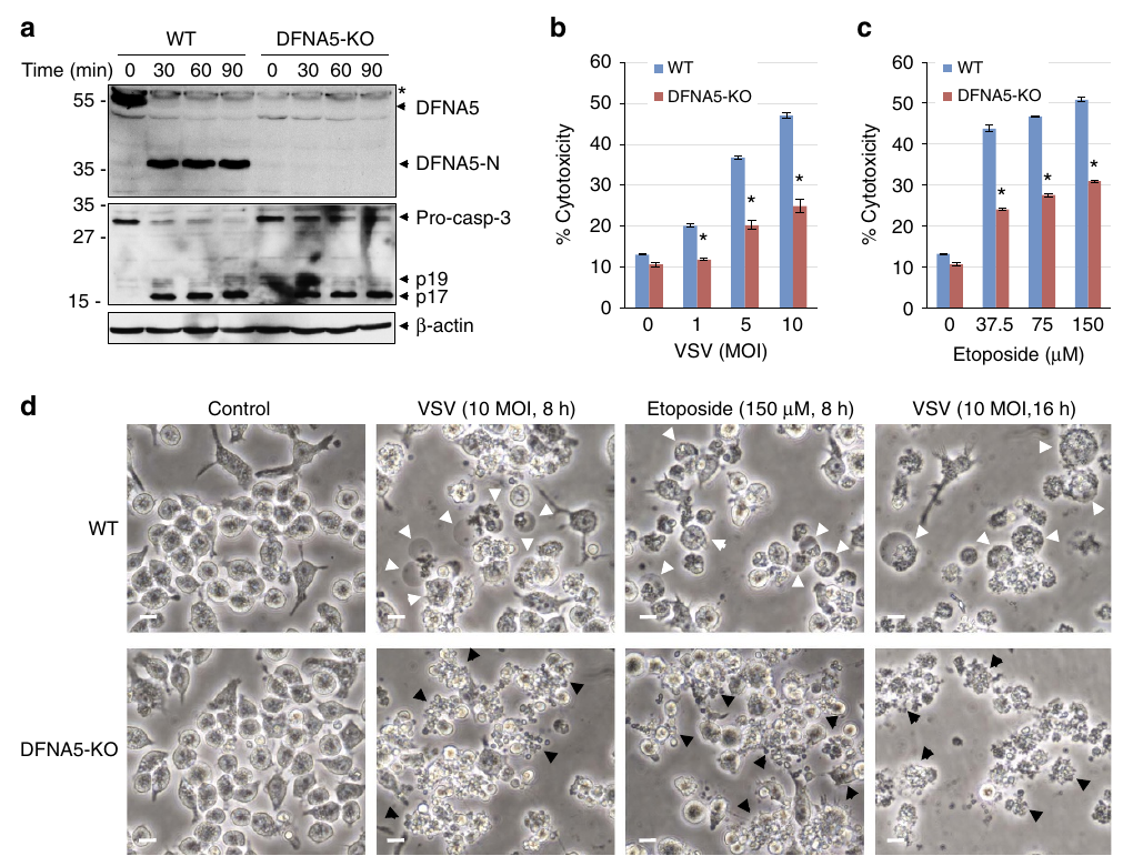
Step 3: Reconnect the mechanism to apoptosis and validate it using knockout mice (Figs. 5–8). The authors induced apoptosis with Bax or active caspase-3 in cells reconstituted with wild-type or D270E-mutant DFNA5. Cells reconstituted with wild-type DFNA5 released large amounts of LDH, whereas those reconstituted with D270E did not undergo membrane rupture. Together, these results indicate that DFNA5, rather than GSDMD, mediates membrane rupture at the end of apoptosis. The authors then validated this in macrophages from DFNA5-knockout mice: secondary necrosis was markedly reduced after VSV infection or etoposide treatment. Time-lapse imaging showed that both cell types initiated apoptosis, but only wild-type cells progressed to membrane rupture.
Conclusion: Secondary necrosis is not passive osmotic disintegration, but a programmed terminal stage executed by the caspase-3–DFNA5 axis. Apoptosis and pyroptosis share the same gasdermin mechanism at the membrane-rupture step.
3. Implications for experiments
- Work backwards from an unusual phenotype to identify the substrate: The starting point was that double-knockout cells still underwent necrosis. "Necroptosis inhibitors have no effect, whereas zVAD works" narrowed the search to non-inflammatory caspases, followed by substrate screening in vitro. This phenotype-driven route to substrate discovery is worth learning from.
- Mutations causing human genetic diseases provide functional clues: Deafness-causing DFNA5 mutations produce a C-terminally truncated protein, suggesting that "the N terminus is cytotoxic and the C terminus controls inhibition". When studying a gene of unknown function, first examine the structural consequences of its disease-causing mutations.
- Consider secondary necrosis when interpreting terminal readouts of apoptosis: LDH release or PI positivity late in an apoptosis experiment may result from programmed membrane rupture mediated by DFNA5. Whether cells express DFNA5/GSDME determines whether the same apoptotic stimulus produces a "quiet" or an "inflammatory" outcome.
4. Connections to other papers on this site
- Extension | GSDMD is the executor of pyroptosis (He 2015, Cell Res): Inflammatory caspases cleave GSDMD to execute pyroptosis, whereas apoptotic caspase-3 cleaves DFNA5 to execute secondary necrosis. The same architecture of "N-terminal killing and C-terminal autoinhibition" serves two cell-death pathways.
- Extension | Gasdermins permeabilize mitochondria to amplify caspase-3 (Rogers 2019, Nat Commun): This paper shows that DFNA5 determines whether apoptotic cells undergo membrane rupture. The 2019 paper further shows that GSDME-N also permeabilizes mitochondria and amplifies caspase-3 activation itself. Together, the two papers provide a complete picture of GSDME function.
- Extension | Caspase-1 initiates apoptosis through Bid (Tsuchiya 2019, Nat Commun): In Tsuchiya et al., secondary necrosis after apoptosis in GSDMD-deficient cells is executed by caspase-3–GSDME, consistent with the mechanism described here.
- Comparison | GSDME releases IL-1β under sublytic conditions (Zhou & Abbott 2021, Cell Rep): This paper identifies GSDME as the executor of secondary necrosis after apoptosis. Zhou and Abbott use endogenous expression levels to show that GSDME normally acts as a sublytic channel for IL-1β release and leads to pyroptosis only when highly expressed. Expression levels determine the outcome of GSDME activity.
(The comparisons above are integrated interpretations across papers, rather than direct conclusions of any single original paper.)
What remains unresolved
Limitations (two points): ① The pore-forming mechanism of DFNA5-N is inferred from homology modelling; pore structures or membrane-lipid binding experiments are not directly shown; ② All evidence comes from cell systems, and the inflammatory consequences of secondary necrosis in vivo were not tested in animal models.
Cleavage of DFNA5 by caspase-3 during apoptosis mediates progression to secondary necrotic/pyroptotic cell death
Rogers, Corey; Fernandes-Alnemri, Teresa; Mayes, Lindsey; Alnemri, Diana; Cingolani, Gino; Alnemri, Emad S. Nature Communications. 2017;8:.
Figures are from the verified original paper stored in the library. Original labels, axes and controls are retained; figure numbers and sources are registered in the library.
References
- Rogers C, Fernandes-Alnemri T, Mayes L, Alnemri D, Cingolani G, Alnemri ES. Cleavage of DFNA5 by caspase-3 during apoptosis mediates progression to secondary necrotic/pyroptotic cell death. Nature Communications. 2017;8:14128. doi:10.1038/ncomms14128
- Wang Y, Gao W, Shi X, et al. Chemotherapy drugs induce pyroptosis through caspase-3 cleavage of a gasdermin. Nature. 2017;547(7661):99-103.
- He WT, Wan H, Hu L, et al. Gasdermin D is an executor of pyroptosis and required for interleukin-1β secretion. Cell Research. 2015;25(12):1285-1298.
- Kayagaki N, Stowe IB, Lee BL, et al. Caspase-11 cleaves gasdermin D for non-canonical inflammasome signalling. Nature. 2015;526(7575):666-671.
Paper discussion
No sign-up. Nickname optional.