- 01
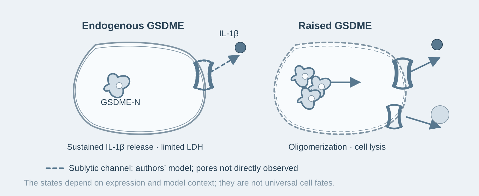
Main finding: After inflammasome activation, endogenous GSDME supports sustained, sublytic (without complete cell membrane rupture) IL-1β release and contributes to membrane leakage even when GSDMD is present;
- 02
Key mechanism: GSDME expression determines the outcome—endogenous levels permit IL-1β release with little lysis, whereas overexpression leads to oligomerization and complete pyroptosis; both gasdermins have a transition period from sublytic to lytic activity, but their kinetics differ;
- 03
Significance: GSDME is a channel for IL-1β release independent of its ability to cause death; in the NLRP1/CARD8 model, both gasdermins are indispensable for IL-1β processing and release.
On this page
1. What question does this paper answer?
The role of GSDME after inflammasome activation has long been disputed: some studies report that it mediates secondary necrosis, while others report that it does nothing. This paper asks: What exactly does GSDME do at endogenous expression levels? The conclusion is that, when GSDMD is absent, inflammasome activation leads to cleavage of GSDME at D270 by apoptotic caspases; endogenous GSDME forms pores that allow IL-1β and small ions to pass without causing substantial cell lysis; only when expression increases does this become complete pyroptosis. GSDME is therefore a channel for IL-1β release whose function is separate from its ability to cause death, and it shares responsibility with GSDMD for IL-1β processing and release in the NLRP1/CARD8 inflammasome.
2. The logic of the evidence
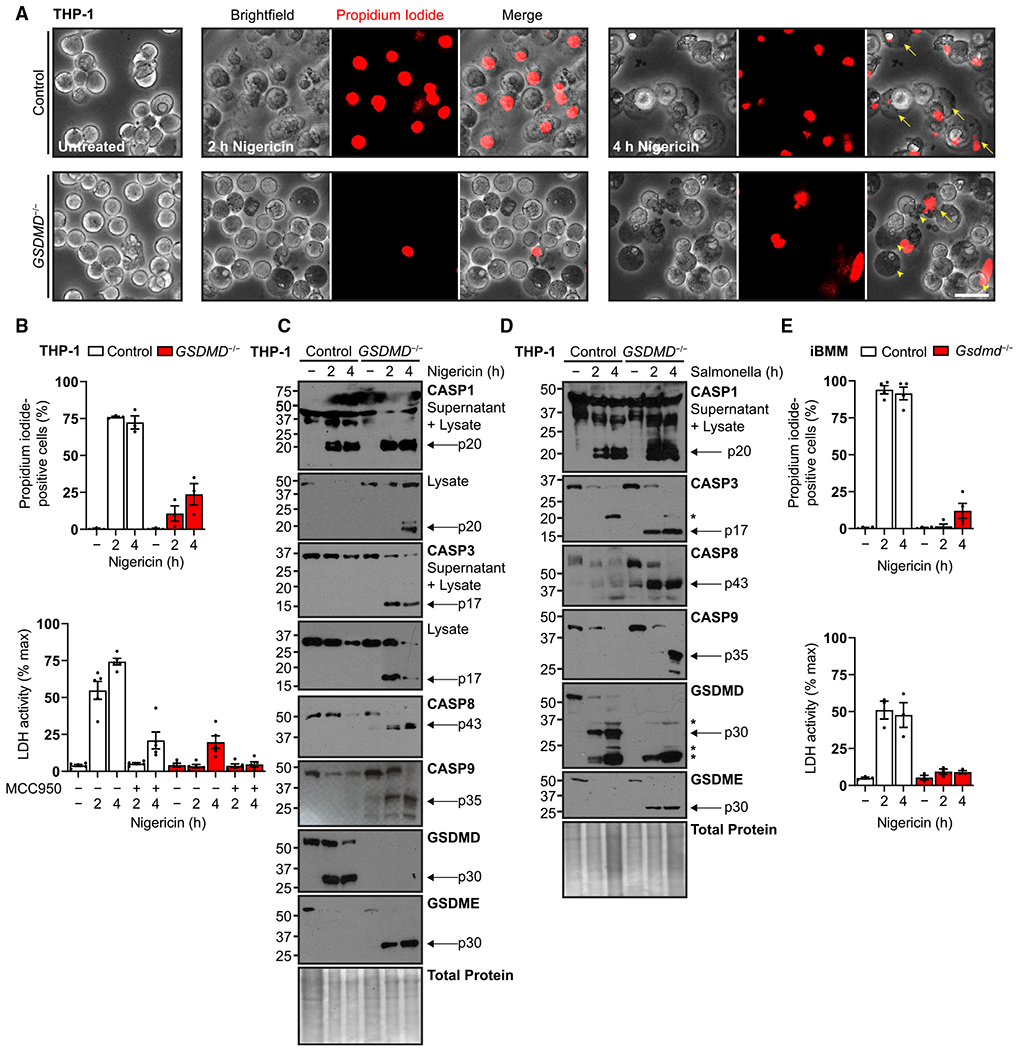
Step 1: When GSDMD is absent, inflammasome signaling switches to the apoptotic caspase–GSDME axis (Fig. 1). The authors activated the NLRP3 (nigericin) or NLRC4 (Salmonella) inflammasome in PMA-differentiated human THP-1 macrophages and found that GSDMD-knockout cells did not undergo typical pyroptosis, but instead displayed apoptotic morphology, caspase-3 activation, and cleavage of GSDME at D270; reconstitution with the non-cleavable GSDME D270A mutant blocked this process. Together, these results indicate that, in the absence of GSDMD, inflammasome signaling switches to GSDME through apoptotic caspase-3.
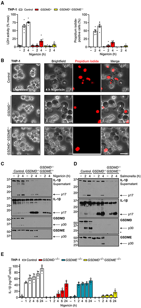
Step 2: Endogenous GSDME permits IL-1β release with essentially no cell lysis (Fig. 2). The authors measured release kinetics in GSDMD-knockout cells and found that IL-1β continued to be released for at least 5 hours, while LDH release (a marker of complete lysis) was limited; in GSDMD/GSDME double-knockout cells, membrane leakage was almost entirely attributable to GSDME. GSDME single knockout did not affect cell death when GSDMD was present. Together, these results indicate that GSDME at endogenous levels forms a sublytic channel for IL-1β release, creating pores that allow small molecules to pass without necessarily killing the cell.
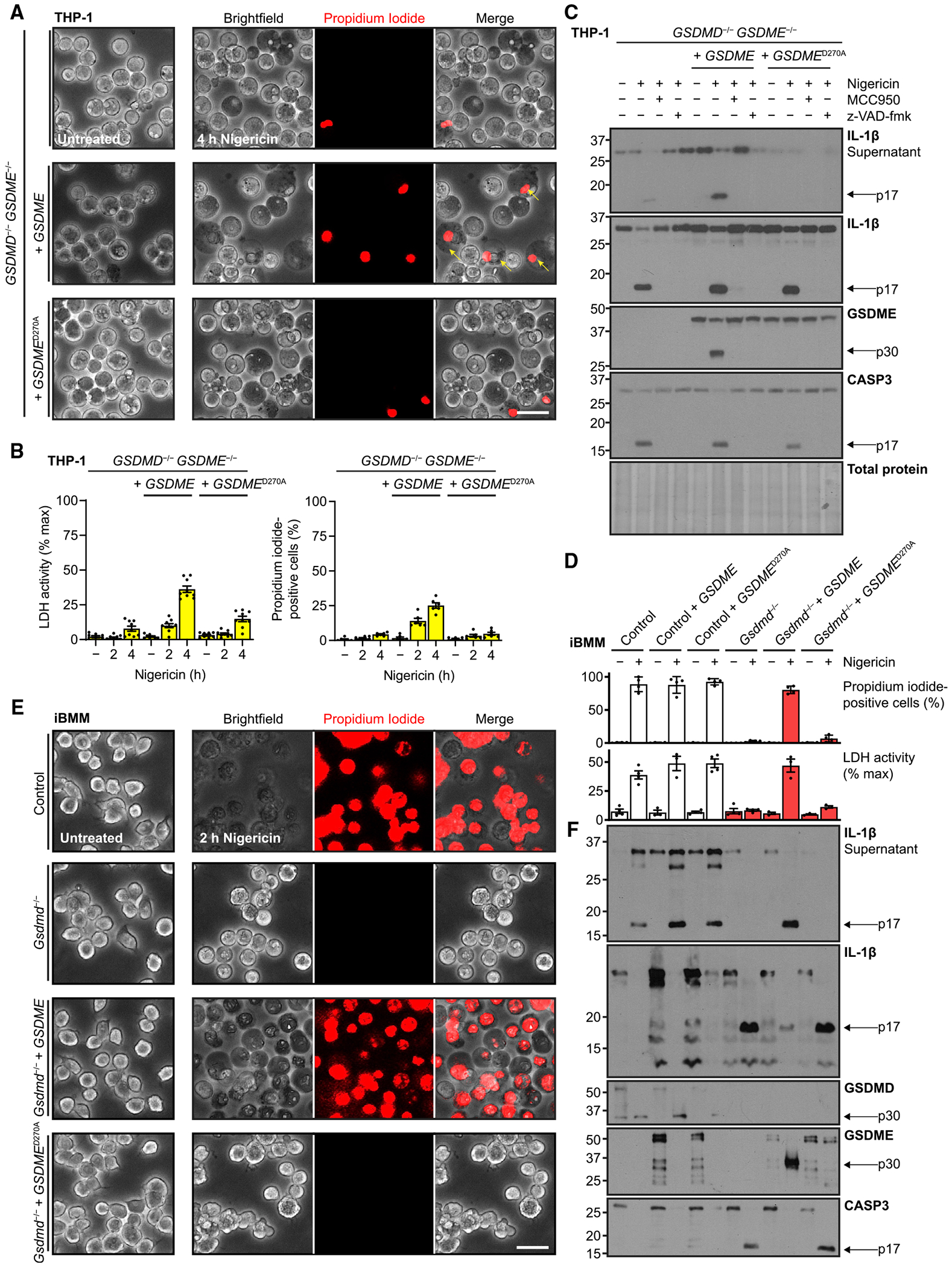
Step 3: Expression level determines sublytic activity versus pyroptosis (Figs. 3–4). The authors reconstituted GSDMD/GSDME double-knockout cells with GSDME at different expression levels and found that reconstitution at endogenous levels supported only sustained release, whereas high expression led to GSDME oligomerization and complete pyroptosis, comparable to GSDMD-mediated pyroptosis. Kinetically, GSDMD-mediated propidium iodide influx peaked at 20 minutes, while LDH release was not complete until 40–60 minutes; GSDME cleavage was nearly complete at 40 minutes, but IL-1β largely remained intracellular during the first hour, with a transition to lytic release at 60–80 minutes. Together, these results indicate that both gasdermins pass through a transition period from sublytic to lytic activity, with different kinetics; whether GSDME causes lysis depends on its expression level.
Step 4: Both gasdermins share responsibility for IL-1β processing and release in the NLRP1/CARD8 model (Fig. 5). The authors treated THP-1 cells with Val-boroPro (a small molecule that inhibits DPP8/9 and activates the NLRP1/CARD8 inflammasome) and found that single knockout of either GSDMD or GSDME disproportionately reduced IL-1β cleavage and output; the pan-caspase inhibitor zVAD completely blocked baseline death in GSDMD-deficient cells without affecting GSDMD-dependent death. Together, these results indicate that in this model, GSDME uncouples IL-1β processing from pyroptotic death, and both gasdermins are indispensable for cytokine output.
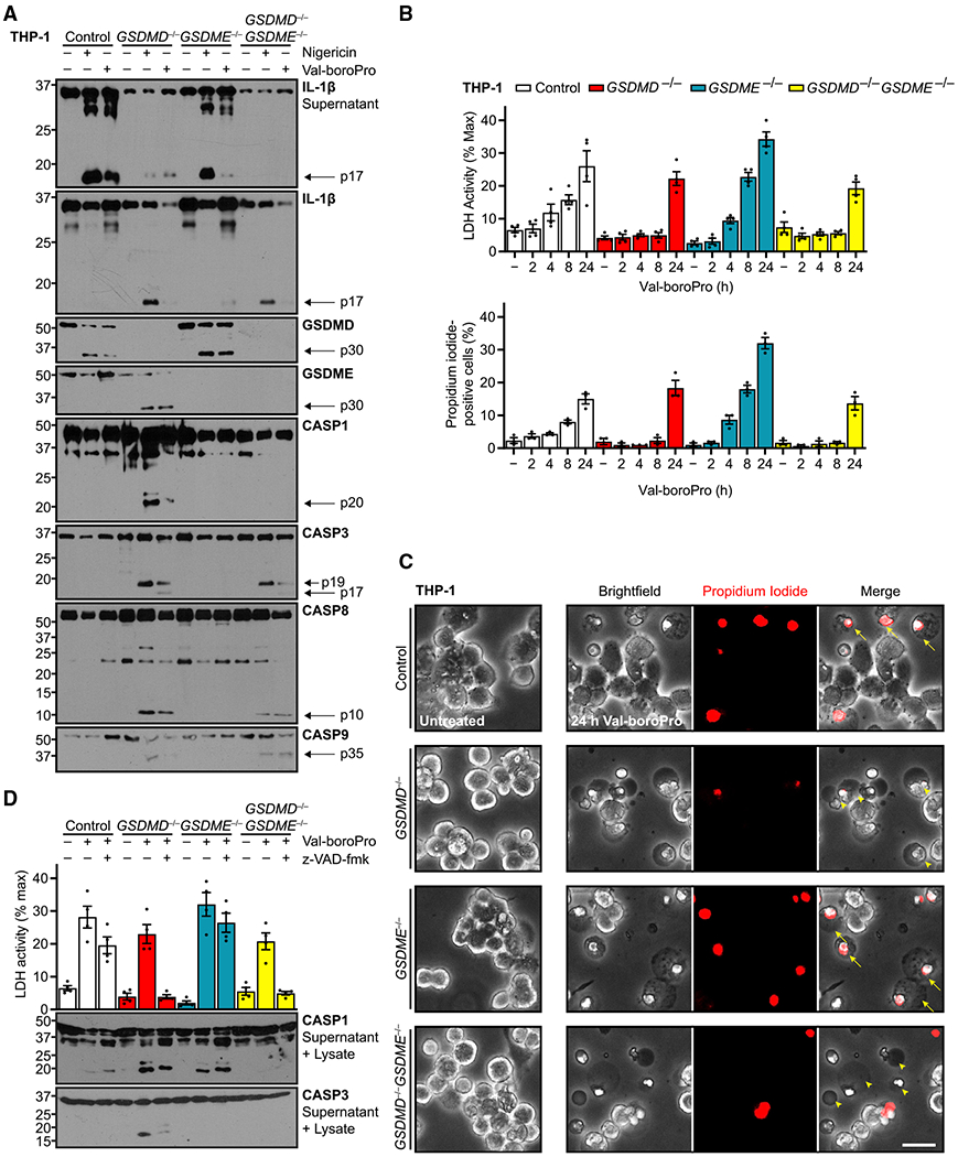
3. Implications for experiments
- Release and lysis should be measured separately: propidium iodide influx, LDH release, and IL-1β detection have different kinetics (approximately 20, 40–60, and 60–80 minutes)—a single readout at a single time point will misclassify "sublytic release" as "no release" or "already lysed."
- Conclusions from GSDME overexpression cannot be extrapolated to endogenous levels: expression in reconstitution experiments should match physiological levels; otherwise, a "sublytic channel" will be interpreted as a "pyroptosis executor."
- Residual death in GSDMD-knockout cells comes from the caspase-3–GSDME axis: when studying GSDMD-specific functions, GSDMD/GSDME double knockout is the clean control.
4. Related papers on this site
- Comparison | GSDME mediates secondary necrosis after apoptosis (Rogers 2017, Nat Commun): Rogers 2017 identifies GSDME as the executor of secondary necrosis after apoptosis; this paper uses endogenous expression levels to demonstrate that the usual role of GSDME is as a sublytic channel for IL-1β release, with pyroptosis occurring only at high expression—expression level determines the outcome of GSDME activity.
- Extension | Incomplete GSDME-mediated pyroptosis permits IL-1α release (Aizawa 2020, iScience): Two "mild" forms of GSDME pores—in this paper, endogenous GSDME acts sublytically, allowing IL-1β release without cell lysis; in Aizawa, when caspase-1 is inhibited, GSDME executes incomplete pyroptosis and permits only IL-1α release—which IL-1 passes through gasdermin pores depends on the upstream caspase.
- Extension | GSDMD permeabilizes mitochondria to amplify pyroptosis (Miao 2023, Immunity): This paper demonstrates that GSDME pores can permit IL-1β release at sublytic levels; Miao demonstrates that GSDMD pores amplify IL-1β release through mitochondrial damage—together, the two papers refine the quantitative relationship between gasdermin pores and IL-1β release.
(The cross-paper comparisons above are a synthesis, not direct conclusions of any single original paper.)
What remains unresolved
Limitations: ① The main conclusions come from the THP-1 cell line and reconstitution systems in immortalized mouse macrophages, with limited validation in primary cells and in vivo; ② the molecular mechanism by which GSDME promotes IL-1β processing remains undefined.
Gasdermin E permits interleukin-1 beta release in distinct sublytic and pyroptotic phases
Bowen Zhou; Derek W. Abbott Cell Reports. 2021;35:108998.
Figures are from the verified original paper stored in the library. Original labels, axes and controls are retained; figure numbers and sources are registered in the library.
References
- Zhou B, Abbott DW. Gasdermin E permits interleukin-1 beta release in distinct sublytic and pyroptotic phases. Cell Rep. 2021;35(2):108998. doi:10.1016/j.celrep.2021.108998
- Rogers C, Erkes DA, Nardone A, et al. Gasdermin pores permeabilize mitochondria to augment caspase-3 activation during apoptosis and inflammasome activation. Nat Commun. 2019;10:1689. doi:10.1038/s41467-019-09397-2
- Tsuchiya K, Nakajima S, Hosojima S, et al. Caspase-1 initiates apoptosis in the absence of gasdermin D. Nat Commun. 2019;10:2091. doi:10.1038/s41467-019-09753-2
- Evavold CL, Ruan J, Tan Y, et al. The pore-forming protein gasdermin D regulates interleukin-1 secretion from living macrophages. Immunity. 2018;48(1):35-44.e6. doi:10.1016/j.immuni.2017.11.013
Paper discussion
No sign-up. Nickname optional.