- 01
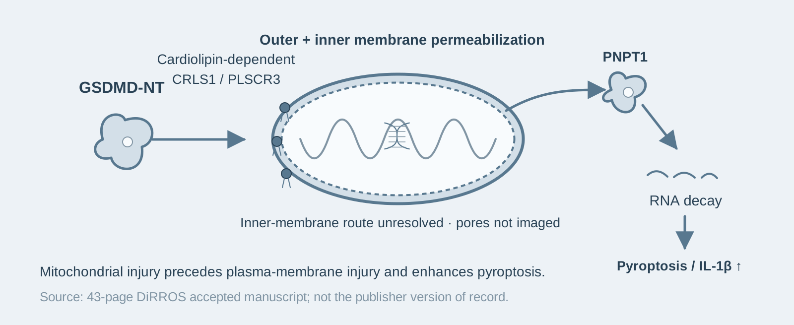
Main finding: During pyroptosis, GSDMD-NT permeabilizes both the inner and outer mitochondrial membranes before the plasma membrane is damaged, leading to fewer mitochondria, loss of membrane potential, increased reactive oxygen species, and release of mtDNA and mitochondrial proteins;
- 02
Key mechanism: The damage depends on GSDMD-NT binding cardiolipin on the outer mitochondrial membrane. Knocking out CRLS1 or PLSCR3 almost eliminates mitochondrial damage; the process does not depend on BAX/BAK or mPTP;
- 03
Significance: Mitochondrial damage is required for pyroptosis and inflammatory cytokine release, and determines pyroptosis-induced antitumor immunity in a tumor model.
On this page
1. What question does this paper answer?
Pyroptosis is known to be accompanied by mitochondrial damage, but is that damage a consequence or a cause? The conclusion of this paper is that the N-terminal pore-forming fragment of GSDMD (GSDMD-NT) permeabilizes both the outer and inner mitochondrial membranes before causing plasma membrane rupture. Mitochondrial contents, including mtDNA and intermembrane-space proteins, are released into the cytosol and, in turn, accelerate and amplify pyroptosis, IL-1β release, and inflammation. All of this depends on cardiolipin on the outer mitochondrial membrane—a phospholipid found mainly in the inner mitochondrial membrane and bacterial membranes—and is independent of the BAX/BAK-mediated mitochondrial permeabilization that occurs during apoptosis.
2. The logic of the evidence
Step 1: Mitochondria are damaged before the plasma membrane during pyroptosis (Figures 1–2). The authors activate canonical (LPS + nigericin) or noncanonical (cytosolic LPS) inflammasomes in immortalized mouse macrophages (iBMDMs). They find that as soon as GSDMD is cleaved, mitochondria show loss of membrane potential, increased reactive oxygen species, decreased oxidative phosphorylation, reduced numbers, and mitophagy, all before plasma membrane damage. Fluorescently tagged GSDMD-NT translocates to mitochondria early in pyroptosis. These results indicate that mitochondrial damage is an early event in pyroptosis, rather than a secondary phenomenon following cell rupture.
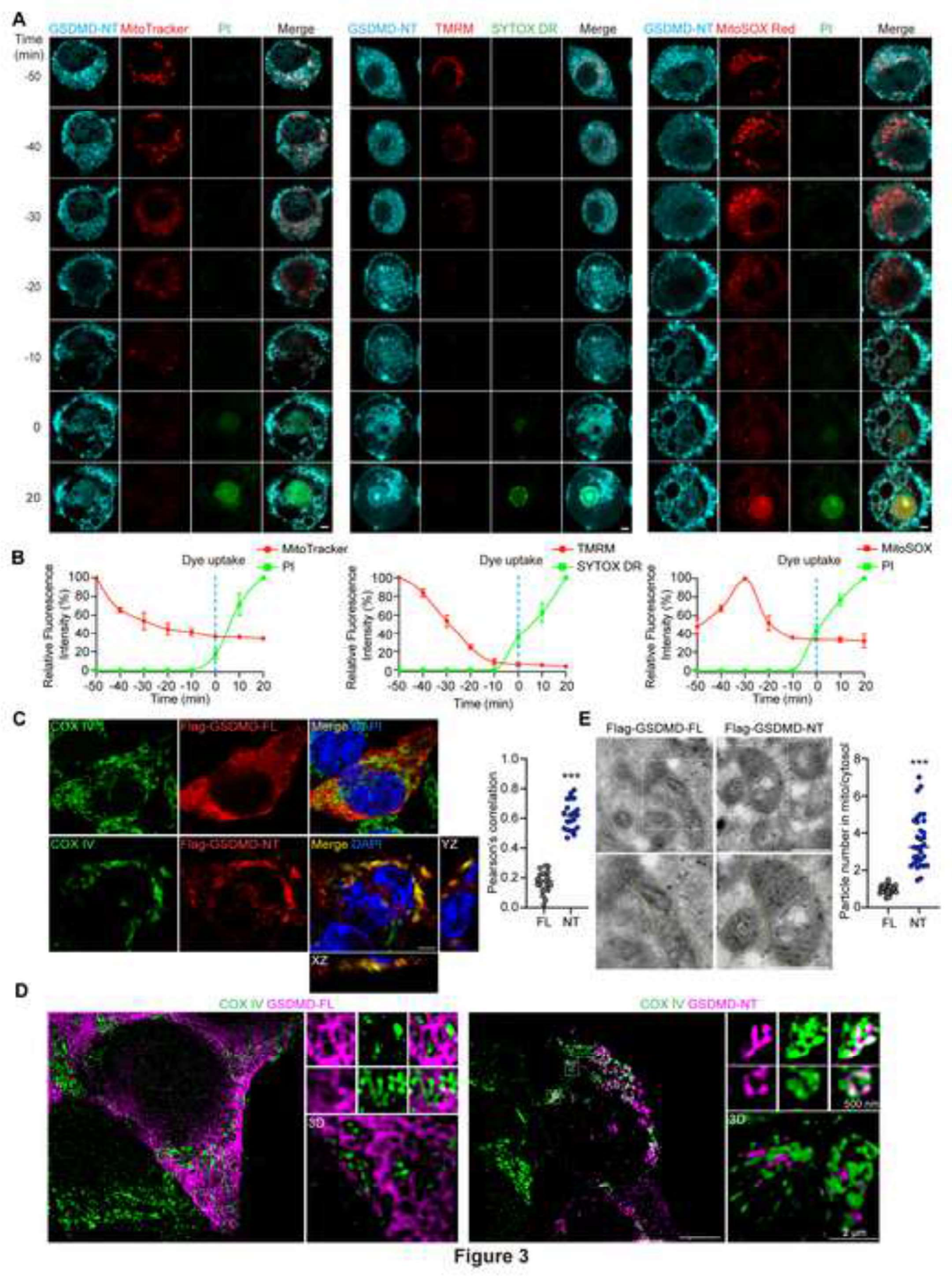
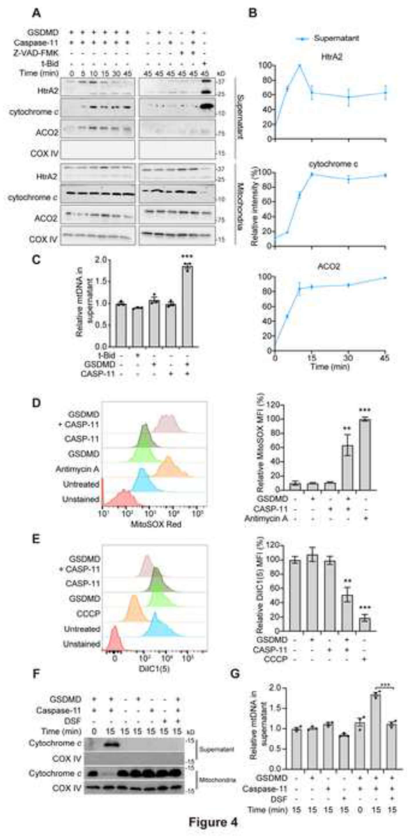
Step 2: Ruling out apoptotic mitochondrial permeabilization (Figure 3). Using BAX/BAK double-knockout cells and treatment with inhibitors of mPTP (the mitochondrial permeability transition pore), the authors find that pyroptotic mitochondrial damage is unaffected. Moreover, whereas apoptosis permeabilizes only the outer membrane (MOMP), pyroptosis permeabilizes both the outer and inner mitochondrial membranes, allowing matrix contents to leak out as well. These results indicate that pyroptotic mitochondrial damage is caused directly by GSDMD-NT and does not proceed through the apoptotic BAX/BAK pathway.
Step 3: Cardiolipin anchors GSDMD-NT-mediated mitochondrial damage (Figures 4–6). GSDMD-NT has a higher affinity for cardiolipin than for plasma membrane phospholipids. The authors incubate recombinant GSDMD-NT with isolated mitochondria in vitro and directly observe permeabilization of both membranes. In cells, knocking out the cardiolipin synthase CRLS1 or the transporter PLSCR3, which flips cardiolipin from the inner to the outer membrane, almost completely eliminates mitochondrial damage. These results indicate that GSDMD-NT permeabilizes mitochondria by binding cardiolipin on the outer membrane; the presence and localization of cardiolipin determine whether damage can occur.
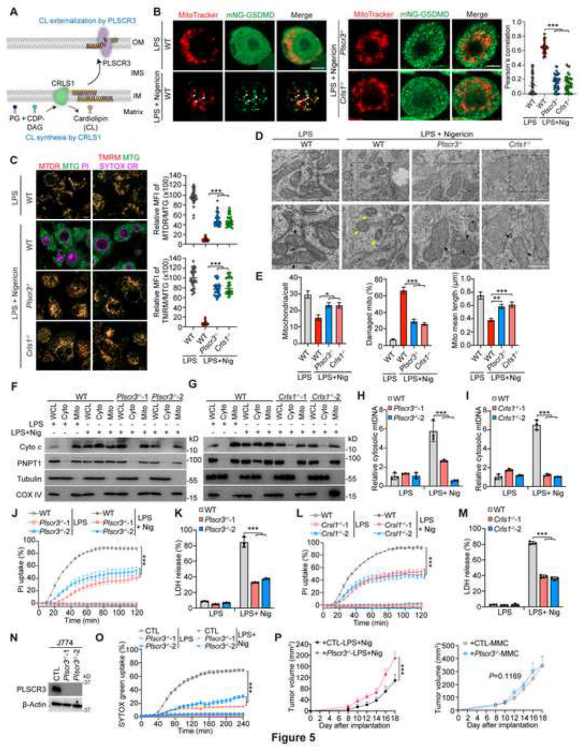
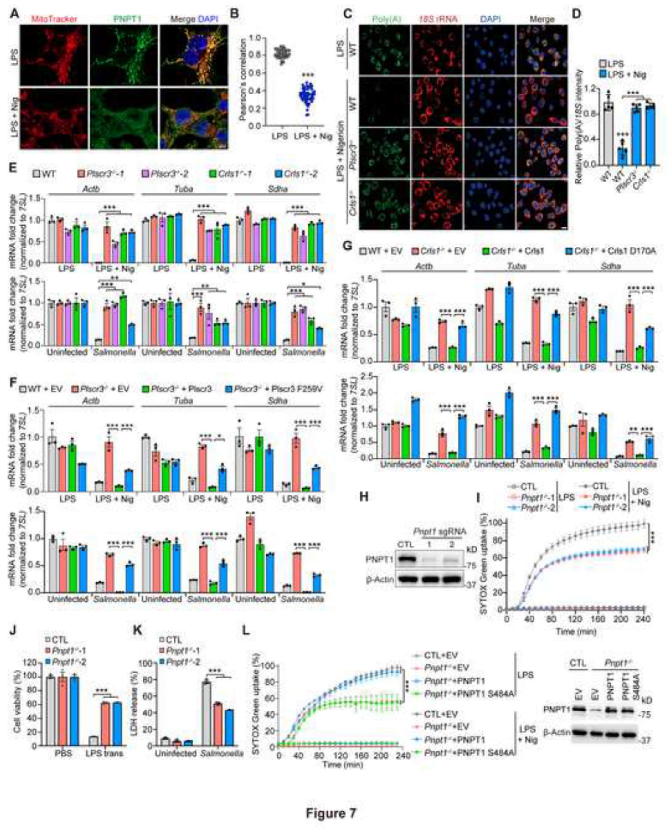
Step 4: The functional consequences of mitochondrial damage (Figures 5–7). In Crls1- or Plscr3-knockout macrophages, both pyroptosis and IL-1β release induced by canonical and noncanonical inflammasomes are markedly inhibited. One mechanism is that permeabilized mitochondria release the intermembrane-space exoribonuclease PNPT1 into the cytosol, causing global mRNA degradation and amplifying pyroptosis. In vivo, the authors use Plscr3-knockout J774 mouse macrophage tumors in a transplantation model and find impaired antitumor immunity induced by tumor-cell pyroptosis and faster tumor growth. This dependence also extends to GSDMA- and GSDME-mediated pyroptosis. These results indicate that mitochondrial damage is required for pyroptosis to proceed fully and for inflammatory amplification, and affects antitumor immunity in vivo.
3. Implications for experiments
- Mitochondrial readouts during pyroptosis are not merely bystander indicators: membrane potential, reactive oxygen species, and mtDNA release in turn determine the intensity of pyroptosis. Monitoring them is equivalent to monitoring an upstream amplifier of pyroptosis.
- BAX/BAK double knockout cannot be used to show that “mitochondria do not participate in pyroptosis”: pyroptotic mitochondrial damage proceeds through the GSDMD-NT–cardiolipin pathway, in which neither BAX/BAK nor mPTP participates. The two mechanisms need to be distinguished using different genetic tools.
- CRLS1/PLSCR3 are genetic handles for modulating the intensity of pyroptosis: when a control is needed that “weakens only the mitochondrial amplification loop without affecting GSDMD cleavage,” these two knockouts provide a cleaner approach than global inhibitors.
4. Related papers on this site
- Further reading | Gasdermin permeabilizes mitochondria to amplify caspase-3 (Rogers 2019, Nat Commun): Rogers 2019 demonstrates that gasdermin-N permeabilizes mitochondria and amplifies caspase-3 through the apoptosome during apoptosis. This paper demonstrates that GSDMD-NT permeabilizes both mitochondrial membranes and amplifies pyroptosis itself in a cardiolipin-dependent manner—the mechanism of “gasdermin attacking mitochondria” serves two cell-death pathways.
- Further reading | Sublytic GSDME-mediated IL-1β release (Zhou & Abbott 2021, Cell Rep): Together, the two papers refine our understanding of “how gasdermin pores quantitatively control IL-1β release.” This paper demonstrates that GSDMD pores amplify release through mitochondrial damage, whereas Zhou & Abbott demonstrate that GSDME pores can permit IL-1β release at a sublytic level without lysing the cell.
(The cross-paper comparisons above are a synthesized interpretation, rather than direct conclusions of either original paper.)
What remains unresolved
Limitations: ① The in vivo evidence is limited to the J774 tumor transplantation model; ② the precise way in which GSDMD-NT permeabilizes the inner membrane—through pores or disruption of the lipid membrane—remains unresolved; ③ some cell types have unknown mechanisms that inhibit mitochondrial damage, which this paper does not systematically identify.
Gasdermin D permeabilization of mitochondrial inner and outer membranes accelerates and enhances pyroptosis
Rui Miao; Cong Jiang; Winston Y. Chang; Haiwei Zhang; Jinsu An; Felicia Ho; Pengcheng Chen; Han Zhang; Caroline Junqueira; Dulguun Amgalan; Felix G. Liang; Junbing Zhang; Charles L. Evavold; Iva Hafner-Bratkovič; Zhibin Zhang; Pietro Fontana; Shiyu Xia; Markus Waldeck-Weiermair; Youdong Pan; Thomas Michel; Liron Bar-Peled; Hao Wu; Jonathan C. Kagan; Richard N. Kitsis; Peng Zhang; Xing Liu; Judy Lieberman Immunity. 2023;56:2523-2541.e8.
The figures are from the author accepted manuscript of Miao et al. (DiRROS, 2023). Complete figures retain their original labels, axes and controls. The manuscript states CC BY-NC-ND without a licence version number; the PDF version, figure numbers and sources are recorded in the library.
References
- Miao R, Jiang C, Chang WY, et al. Gasdermin D permeabilization of mitochondrial inner and outer membranes accelerates and enhances pyroptosis. Immunity. 2023;56(11):2523-2541.e8. doi:10.1016/j.immuni.2023.10.004
- Rogers C, Erkes DA, Nardone A, et al. Gasdermin pores permeabilize mitochondria to augment caspase-3 activation during apoptosis and inflammasome activation. Nat Commun. 2019;10:1689. doi:10.1038/s41467-019-09397-2
- Ding J, Wang K, Liu W, et al. Pore-forming activity and structural autoinhibition of the gasdermin family. Nature. 2016;535(7610):111-116. doi:10.1038/nature18590
Paper discussion
No sign-up. Nickname optional.